1CVS image
Deposition Date 1999-08-24
Release Date 2000-01-28
Last Version Date 2024-11-20
Entry Detail
PDB ID:
1CVS
Title:
CRYSTAL STRUCTURE OF A DIMERIC FGF2-FGFR1 COMPLEX
Biological Source:
Source Organism:
Homo sapiens (Taxon ID: 9606)
Host Organism:
Method Details:
Experimental Method:
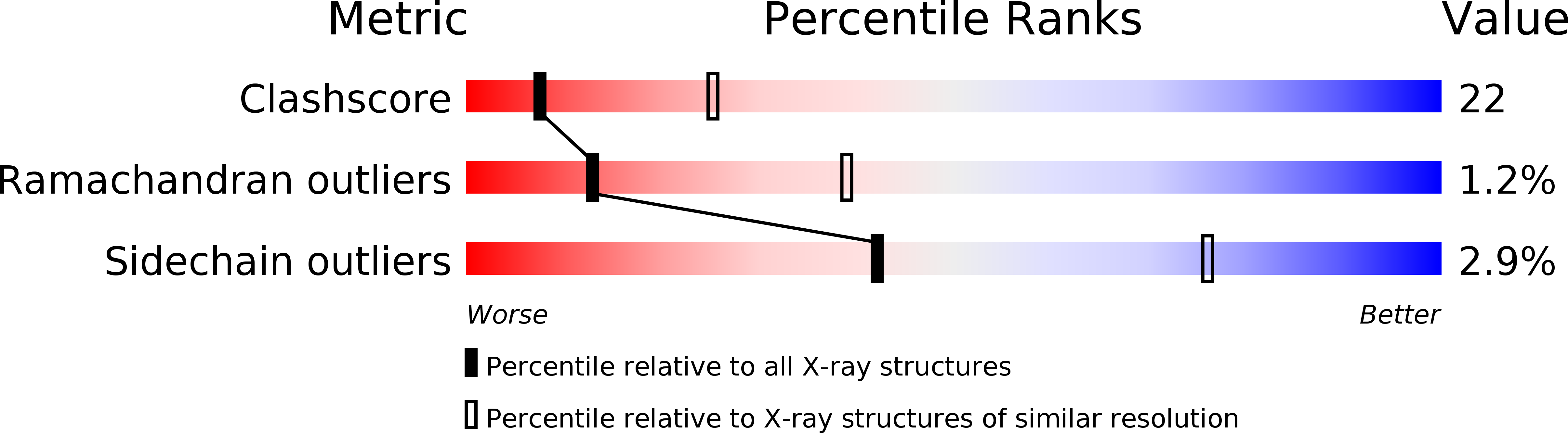
Resolution:
2.80 Å
R-Value Free:
0.28
R-Value Work:
0.24
R-Value Observed:
0.24
Space Group:
P 41 21 2
Macromolecular Entities
Structures with similar UniProt ID
Protein Blast
Polymer Type:polypeptide(L)
Molecule:FIBROBLAST GROWTH FACTOR 2
Gene (Uniprot):FGF2
Mutagens:YES
Chain IDs:A, B
Chain Length:132
Number of Molecules:2
Biological Source:Homo sapiens
Structures with similar UniProt ID
Protein Blast
Polymer Type:polypeptide(L)
Molecule:FIBROBLAST GROWTH FACTOR RECEPTOR 1
Gene (Uniprot):FGFR1
Mutagens:YES
Chain IDs:C, D
Chain Length:225
Number of Molecules:2
Biological Source:Homo sapiens
Ligand Molecules
Primary Citation
Structural basis for FGF receptor dimerization and activation.
Cell(Cambridge,Mass.) 98 641 650 (1999)
PMID: 10490103 DOI: 10.1016/S0092-8674(00)80051-3

Abstact

The crystal structure of FGF2 bound to a naturally occurring variant of FGF receptor 1 (FGFR1) consisting of immunoglobulin-like domains 2 (D2) and 3 (D3) has been determined at 2.8 A resolution. Two FGF2:FGFR1 complexes form a 2-fold symmetric dimer. Within each complex, FGF2 interacts extensively with D2 and D3 as well as with the linker between the two domains. The dimer is stabilized by interactions between FGF2 and D2 of the adjoining complex and by a direct interaction between D2 of each receptor. A positively charged canyon formed by a cluster of exposed basic residues likely represents the heparin-binding site. A general model for FGF- and heparin-induced FGFR dimerization is inferred from the crystal structure, unifying a wealth of biochemical data.

Legend

Protein

Chemical

Disease

Primary Citation of related structures