1CUR image
Deposition Date 1996-04-19
Release Date 1996-11-08
Last Version Date 2024-05-22
Entry Detail
PDB ID:
1CUR
Title:
REDUCED RUSTICYANIN, NMR
Biological Source:
Source Organism:
Method Details:
Experimental Method:
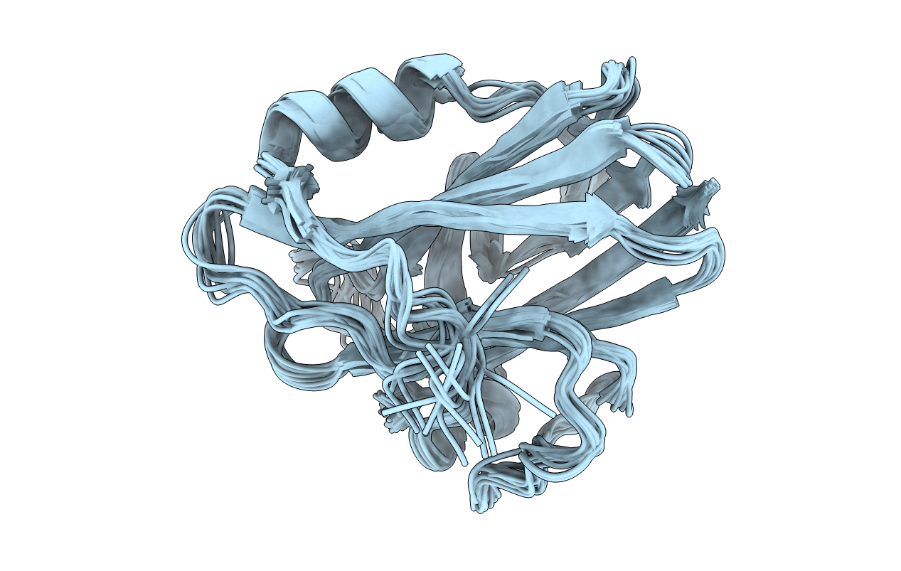
Conformers Submitted:
15
Macromolecular Entities
Polymer Type:polypeptide(L)
Molecule:CU(I) RUSTICYANIN
Chain IDs:A
Chain Length:155
Number of Molecules:1
Biological Source:Acidithiobacillus ferrooxidans
Ligand Molecules
Primary Citation
NMR solution structure of Cu(I) rusticyanin from Thiobacillus ferrooxidans: structural basis for the extreme acid stability and redox potential.
J.Mol.Biol. 263 752 767 (1996)
PMID: 8947573 DOI: 10.1006/jmbi.1996.0613

Abstact

The solution structure of the Cu(I) form of the rusticyanin from Thiobacillus ferrooxidans has been calculated from a total of 1979 distance and dihedral angle constraints derived from 1H, 13C and 15N NMR spectra. The structures reveal two beta-sheets, one of six strands and one of seven strands that are tightly packed in a beta-barrel or beta-sandwich arrangement, and a short helix that extends on the outside of one of the sheets to form a second hydrophobic core. The copper coordination sphere is composed of the standard type I ligands (His2CysMet) in a distorted tetrahedral arrangement. The copper-binding site is located within a hydrophobic region at one end of the molecule, surrounded by a number of aromatic rings and hydrophobic residues. This configuration probably contributes to the acid stability of the copper site, since close association of the aromatic rings with the histidine ligands would sterically hinder their dissociation from the copper. An electrostatic analysis based on a comparison of the structures of rusticyanin and French bean plastocyanin shows that factors determining the high redox potential of rusticyanin include contributions from charged side-chains and from the disposition of backbone peptide dipoles, particularly in the 81 to 86 region of the sequence and the ligand cysteine residue. These interactions should also contribute to the acid stability by inhibiting protonation of His143.

Legend

Protein

Chemical

Disease

Primary Citation of related structures