1CUN image
Deposition Date 1999-08-20
Release Date 1999-10-06
Last Version Date 2024-02-07
Entry Detail
PDB ID:
1CUN
Title:
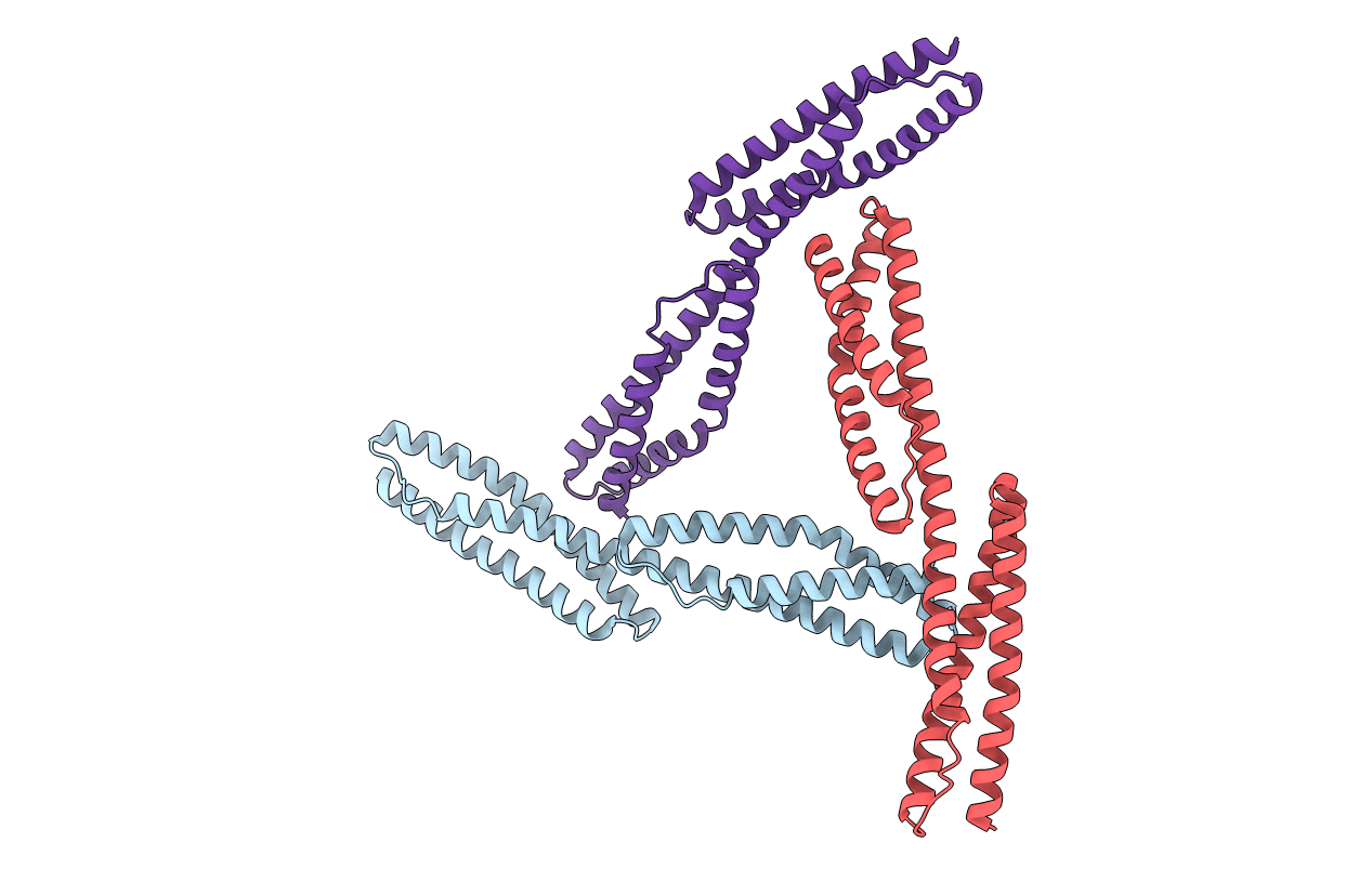
CRYSTAL STRUCTURE OF REPEATS 16 AND 17 OF CHICKEN BRAIN ALPHA SPECTRIN
Biological Source:
Source Organism:
Gallus gallus (Taxon ID: 9031)
Method Details:
Experimental Method:
Resolution:
2.00 Å
R-Value Free:
0.25
R-Value Work:
0.22
Space Group:
C 2 2 21
Macromolecular Entities
Structures with similar UniProt ID
Protein Blast
Polymer Type:polypeptide(L)
Molecule:PROTEIN (ALPHA SPECTRIN)
Gene (Uniprot):SPTAN1
Mutagens:L1771V, G1827S
Chain IDs:A, B, C
Chain Length:213
Number of Molecules:3
Biological Source:Gallus gallus
Primary Citation
Structures of two repeats of spectrin suggest models of flexibility.
Cell(Cambridge,Mass.) 98 523 535 (1999)
PMID: 10481916 DOI: 10.1016/S0092-8674(00)81980-7

Abstact

Spectrin is a vital component of the cytoskeleton, conferring flexibility on cells and providing a scaffold for a variety of proteins. It is composed of tandem, antiparallel coiled-coil repeats. We report four related crystal structures at 1.45 A, 2.0 A, 3.1 A, and 4.0 A resolution of two connected repeats of chicken brain alpha-spectrin. In all of the structures, the linker region between adjacent units is alpha-helical without breaks, kinks, or obvious boundaries. Two features observed in the structures are (1) conformational rearrangement in one repeat, resulting in movement of the position of a loop, and (2) varying degrees of bending at the linker region. These features form the basis of two different models of flexibility: a conformational rearrangement and a bending model. These models provide novel atomic details of spectrin flexibility.

Legend

Protein

Chemical

Disease

Primary Citation of related structures