Deposition Date
1999-08-20
Release Date
2000-04-17
Last Version Date
2024-10-30
Entry Detail
PDB ID:
1CU4
Keywords:
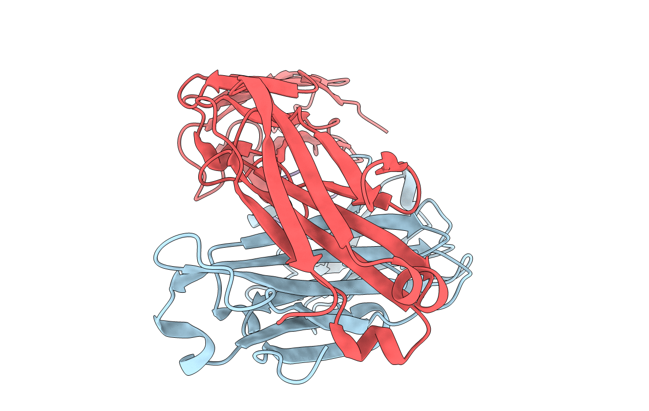
Title:
CRYSTAL STRUCTURE OF THE ANTI-PRION FAB 3F4 IN COMPLEX WITH ITS PEPTIDE EPITOPE
Biological Source:
Source Organism(s):
Mus musculus (Taxon ID: 10090)
Method Details:
Experimental Method:
Resolution:
2.90 Å
R-Value Free:
0.26
R-Value Work:
0.15
Space Group:
P 21 21 21