1CTA image
Deposition Date 1992-11-12
Release Date 1993-10-31
Last Version Date 2024-10-23
Entry Detail
PDB ID:
1CTA
Keywords:
Title:
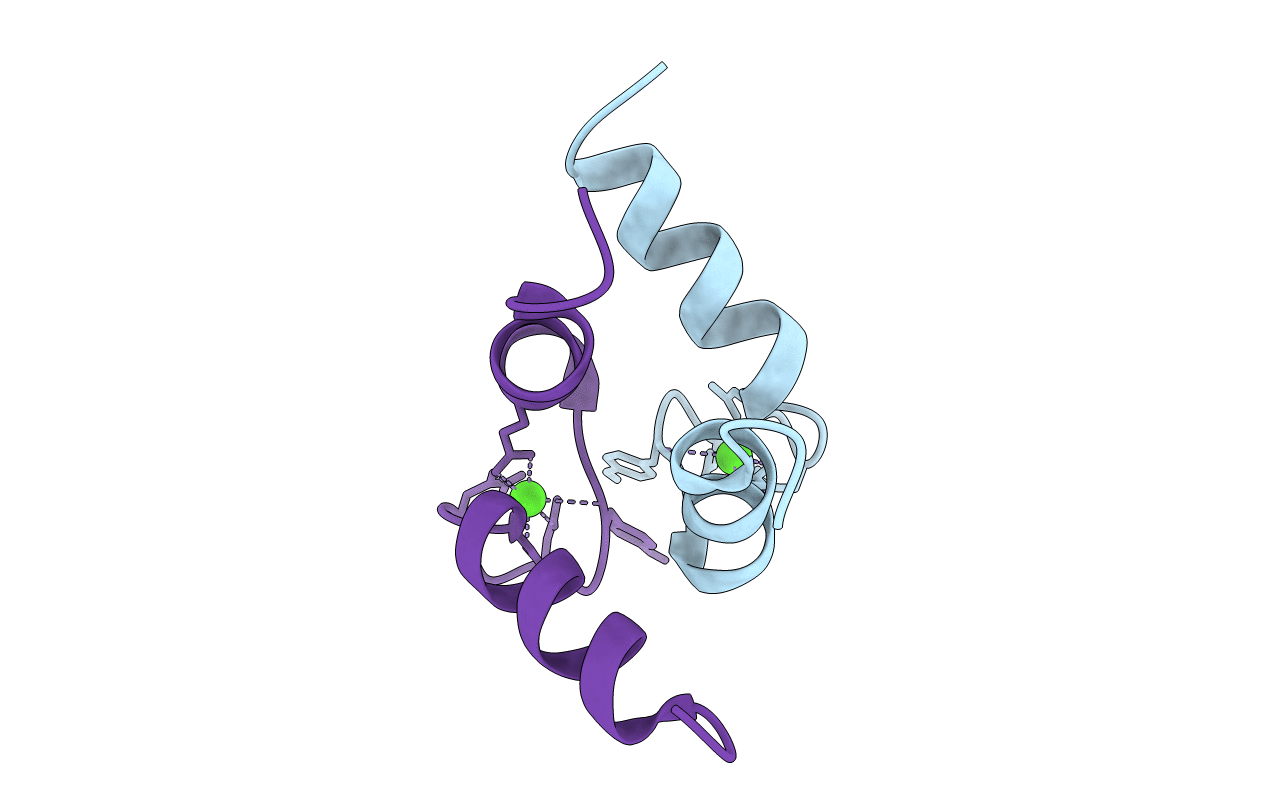
DETERMINATION OF THE SOLUTION STRUCTURE OF A SYNTHETIC TWO-SITE CALCIUM-BINDING HOMODIMERIC PROTEIN DOMAIN BY NMR SPECTROSCOPY
Biological Source:
Source Organism:
(Taxon ID: ) (Taxon ID: )
Method Details:
Experimental Method:
Conformers Submitted:
1
Macromolecular Entities
Polymer Type:polypeptide(L)
Molecule:TROPONIN C SITE III - SITE III HOMODIMER
Gene (Uniprot):TNNC2
Chain IDs:A, B
Chain Length:36
Number of Molecules:2
Biological Source:
Ligand Molecules
Primary Citation
Determination of the solution structure of a synthetic two-site calcium-binding homodimeric protein domain by NMR spectroscopy.
Biochemistry 31 9572 9580 (1992)
PMID: 1390738 DOI: 10.1021/bi00155a009

Abstact

The solution structure of a 34-residue synthetic calcium-binding peptide from site III of chicken troponin-C has been determined by 1H NMR spectroscopy. In solution and in the presence of calcium this peptide forms a symmetric two-site homodimeric calcium-binding domain (Shaw et al., 1990). The solution structure of this dimer was determined from the measurement of 470 NOEs from a 75-ms NOESY data set. For the dimer structure determination, the constraint list included 868 distance restraints, 44 phi angles, and 24 chi 1 and 2 chi 2 angles. Seven structures were calculated by restrained molecular dynamics using a procedure in which intramonomer distances were used first and then all distances, intra- and intermonomer, were input during further dynamics. The structures exhibited a fold very similar to the C-terminal domain of troponin-C comprised of a pair of helix-loop-helix calcium-binding sites. The rms deviation of these structures for backbone atoms between residues 97-122 and 97'-122' for the dimer was 0.82 A. The dimer structure was also calculated to be more symmetric than sites III and IV in troponin-C.

Legend

Protein

Chemical

Disease

Primary Citation of related structures