
Deposition Date
1999-08-20
Release Date
1999-12-15
Last Version Date
2024-02-07
Entry Detail
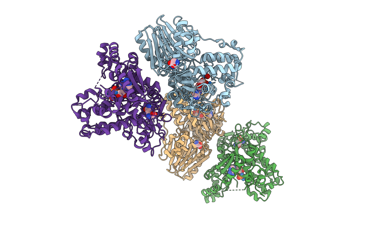
PDB ID:
1CT9
Keywords:
Title:
CRYSTAL STRUCTURE OF ASPARAGINE SYNTHETASE B FROM ESCHERICHIA COLI
Biological Source:
Source Organism(s):
Escherichia coli (Taxon ID: 562)
Expression System(s):
Method Details:
Experimental Method:
Resolution:
2.00 Å
R-Value Free:
0.29
R-Value Work:
0.19
R-Value Observed:
0.19
Space Group:
P 21 21 21
