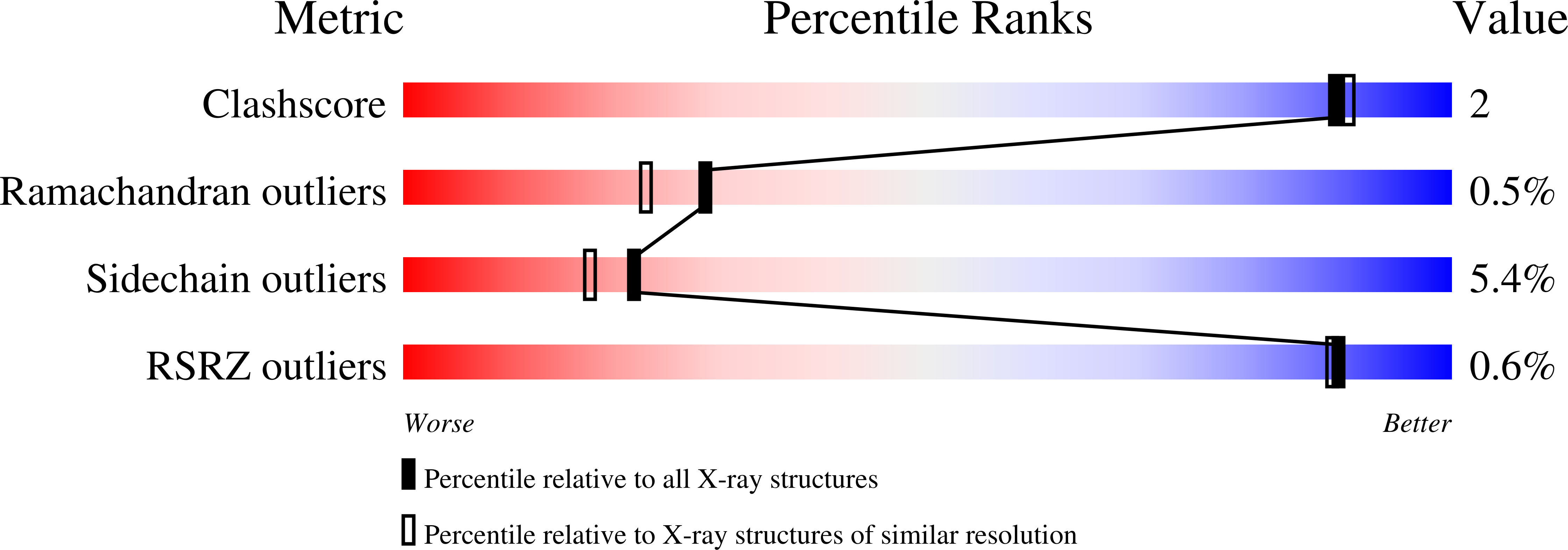
1CRW image
Deposition Date 1999-08-16
Release Date 2000-09-20
Last Version Date 2024-02-07
Entry Detail
PDB ID:
1CRW
Keywords:
Title:
CRYSTAL STRUCTURE OF APO-GLYCERALDEHYDE-3-PHOSPHATE DEHYDROGENASE FROM PALINURUS VERSICOLOR AT 2.0A RESOLUTION
Biological Source:
Source Organism:
Method Details:
Experimental Method:
Resolution:
2.00 Å
R-Value Free:
0.22
R-Value Work:
0.16
Space Group:
C 1 2 1
Macromolecular Entities
Structures with similar UniProt ID
Protein Blast
Polymer Type:polypeptide(L)
Molecule:D-GLYCERALDEHYDE-3-PHOSPHATE-DEHYDROGENASE
Chain IDs:A (auth: G), B (auth: R)
Chain Length:333
Number of Molecules:2
Biological Source:Palinurus versicolor
Primary Citation
Structure of apo-glyceraldehyde-3-phosphate dehydrogenase from Palinurus versicolor.
J.Struct.Biol. 130 1 9 (2000)
PMID: 10806086 DOI: 10.1006/jsbi.2000.4220

Abstact

d-Glyceraldehyde-3-phosphate dehydrogenase (GAPDH) shows cooperative properties for binding coenzymes. The structure of apo-GAPDH from Palinurus versicolor has been solved at 2.0 A resolution by X-ray crystallography. The final model gives a crystallographic R factor of 0.178 in the resolution range 8 to 2 A. The structural comparison with holo-GAPDH from the same species reveals a conformational change induced by coenzyme binding similar to that observed in Bacillus stearothermophilus GAPDH but to a lesser extent. The differences in magnitude during the apo-holo transition between these two enzymes were analyzed with respect to the change of the amino acid composition in the coenzyme binding pocket. In the crystalline state of apo-GAPDH, the overall structures of the subunits are similar to each other; however, significant differences in temperature factors and minor differences in domain rotation upon coenzyme binding were observed for different subunits. These structural features are discussed in relation to the environmental asymmetry of crystallographically independent subunits.

Legend

Protein

Chemical

Disease

Primary Citation of related structures