
Deposition Date
1994-05-11
Release Date
1995-01-26
Last Version Date
2024-11-06
Entry Detail
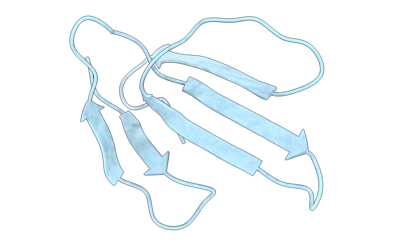
PDB ID:
1COD
Keywords:
Title:
SOLUTION CONFORMATION OF COBROTOXIN: A NUCLEAR MAGNETIC RESONANCE AND HYBRID DISTANCE GEOMETRY-DYNAMICAL SIMULATED ANNEALING STUDY
Biological Source:
Method Details:
Experimental Method:
Conformers Submitted:
1
