1COC image
Deposition Date 1999-05-26
Release Date 1999-06-09
Last Version Date 2023-12-27
Entry Detail
PDB ID:
1COC
Keywords:
Title:
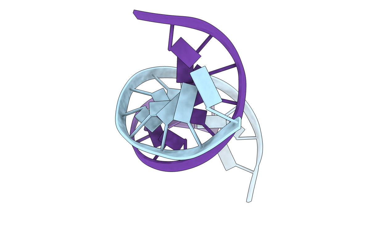
SOLUTION-STATE STRUCTURE OF A DNA DODECAMER DUPLEX CONTAINING A CIS-SYN THYMINE CYCLOBUTANE DIMER.
Biological Source:
Source Organism:
(Taxon ID: ) (Taxon ID: )
Method Details:
Experimental Method:
Conformers Submitted:
1
Macromolecular Entities
Polymer Type:polydeoxyribonucleotide
Molecule:DNA (5'-D(*GP*CP*AP*CP*GP*AP*AP*TP*TP*AP*AP*G)-3')
Chain IDs:A
Chain Length:12
Number of Molecules:1
Biological Source:
Polymer Type:polydeoxyribonucleotide
Molecule:DNA (5'-D(*CP*TP*TP*AP*AP*TP*TP*CP*GP*TP*GP*C)-3')
Chain IDs:B
Chain Length:12
Number of Molecules:1
Biological Source:
Ligand Molecules
Primary Citation
Solution-state structure of a DNA dodecamer duplex containing a Cis-syn thymine cyclobutane dimer, the major UV photoproduct of DNA.
J.Mol.Biol. 282 1013 1032 (1998)
PMID: 9753551 DOI: 10.1006/jmbi.1998.2062

Abstact

The solution structures of a duplex DNA dodecamer containing a cis-syn cyclobutane thymine dimer d(GCACGAAT[cs]TAAG).d(CTTAATTCG TGC) and its native parent sequence were determined using NMR data collected at 750 MHz. The dodecamer sequence corresponds to the section of a site-specific cis-syn dimer containing 49-mer that was found to be the binding site for the dimer-specific T4 denV endonuclease V repair enzyme by chemical and enzymatic footprinting experiments. Structures of both sequences were derived from NOE restrained molecular dynamics/simulated annealing calculations using a fixed outer layer of water and an inner dynamic layer of water with sodium counterions. The resulting structures reveal a subtle distortion to the phosphodiester backbone in the dimer-containing sequence which includes a BII phosphate at the T9pA10 junction immediately 3' to the dimer. The BII phosphate, established experimentally by analysis of the 31P chemical shifts and interpretation of the 3JP-H3' values using an optimized Karplus relationship, enables the DNA helix to accommodate the dimer by destacking the base 3' to the dimer. Furthermore, the structures provide explanations for the unusually shifted T8-N3H imino, A16-H2 and T8-Me proton resonances and T9pA10 (31)P NMR resonance and are consistent with bending, unwinding, and thermodynamic data. The implications of the structural data for the mechanism by which cis-syn dimers are recognized by repair enzymes and bypassed by DNA polymerases are also discussed.

Legend

Protein

Chemical

Disease

Primary Citation of related structures