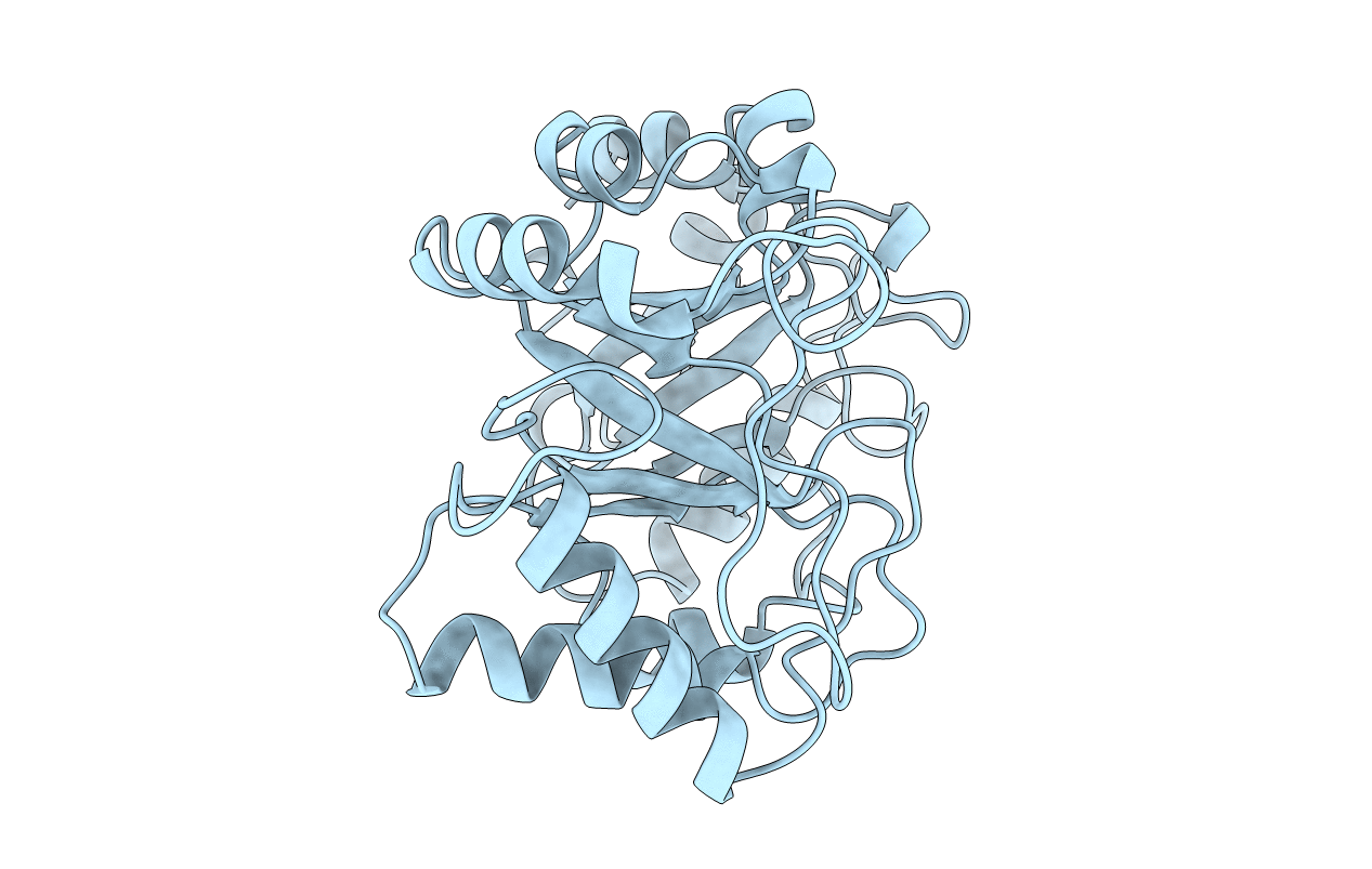
1CNV image
Deposition Date 1995-02-20
Release Date 1996-03-08
Last Version Date 2024-10-16
Entry Detail
PDB ID:
1CNV
Keywords:
Title:
CRYSTAL STRUCTURE OF CONCANAVALIN B AT 1.65 A RESOLUTION
Biological Source:
Source Organism:
Method Details:
Experimental Method:
Resolution:
1.65 Å
R-Value Work:
0.17
R-Value Observed:
0.17
Space Group:
P 61
Macromolecular Entities
Structures with similar UniProt ID
Protein Blast
Polymer Type:polypeptide(L)
Molecule:CONCANAVALIN B
Chain IDs:A
Chain Length:299
Number of Molecules:1
Biological Source:Canavalia ensiformis
Primary Citation
Crystal structure of concanavalin B at 1.65 A resolution. An "inactivated" chitinase from seeds of Canavalia ensiformis.
J.Mol.Biol. 254 237 246 (1995)
PMID: 7490746 DOI: 10.1006/jmbi.1995.0614

Abstact

Seeds of Canavalia ensiformis (jack bean) contain besides large amounts of canavalin and concanavalin A, a protein with a molecular mass of 33,800 which has been named concanavalin B. Although concanavalin B shares about 40% sequence identity with plant chitinases belonging to glycosyl hydrolase family 18, no chitinase activity could be detected for this protein. To resolve this incongruity concanavalin B was crystallised and its three-dimensional structure determined at 1.65 A (1 A = 0.1 nm) resolution. The structure consists of a single domain with a (beta/alpha)8 topology. A 30 amino acid residue long loop occurs between the second beta-strand of the barrel and the second alpha-helix. This extended loop is unusual for the (beta/alpha)8 topology, but appears in a similar conformation in the structures of the seed protein narbonin and several chitinases as well. Two non-proline cis-peptide bonds are present in the structure of concanavalin B: Ser34-Phe, and Trp265-Asn. This structural feature is rarely observed in proteins, but could also be identified in the three-dimensional structures of family 18 chitinases and narbonin in coincident positions. In the chitinases the aromatic residues of the non-proline cis-peptides have been proposed to have a function in the binding of the substrate. The region in concanavalin B, where in chitinases the active site is located, shows two significant differences. First, the catalytic glutamic acid is a glutamine in concanavalin B. Second, although part of the substrate binding cleft of the chitinases is present in concanavalin B, it is much shorter. From this we conclude that concanavalin B and family 18 chitinases are closely related, but that concanavalin B has lost its enzymatic function. It still may act as a carbohydrate binding protein, however.

Legend

Protein

Chemical

Disease

Primary Citation of related structures
Feedback Form
Name
Email
Institute
Feedback