1CMX image
Deposition Date 1999-05-12
Release Date 1999-07-27
Last Version Date 2023-12-27
Entry Detail
PDB ID:
1CMX
Keywords:
Title:
STRUCTURAL BASIS FOR THE SPECIFICITY OF UBIQUITIN C-TERMINAL HYDROLASES
Biological Source:
Source Organism:
(Taxon ID: )
Method Details:
Experimental Method:
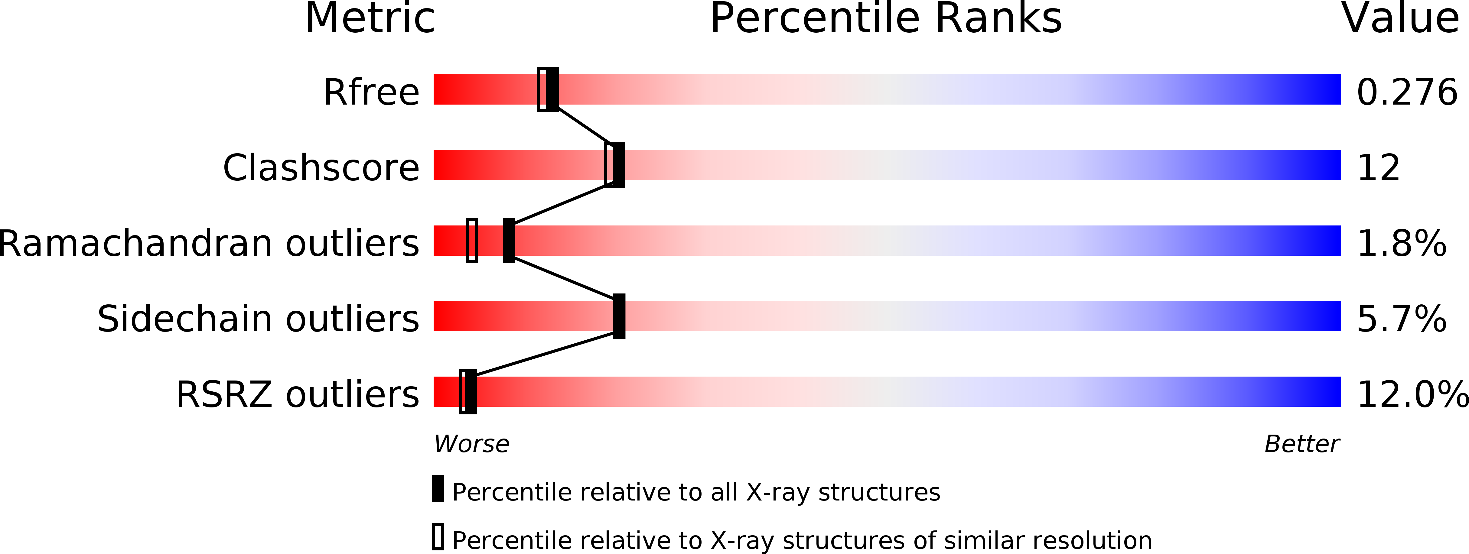
Resolution:
2.25 Å
R-Value Free:
0.28
R-Value Work:
0.24
Space Group:
H 3
Macromolecular Entities
Polymer Type:polypeptide(L)
Molecule:PROTEIN (UBIQUITIN YUH1-UBAL)
Gene (Uniprot):YUH1
Chain IDs:A, C
Chain Length:235
Number of Molecules:2
Biological Source:
Polymer Type:polypeptide(L)
Molecule:PROTEIN (UBIQUITIN YUH1-UBAL)
Chain IDs:B, D
Chain Length:76
Number of Molecules:2
Biological Source:
Modified Residue
Compound ID Chain ID Parent Comp ID Details 2D Image
GLZ B GLY AMINO-ACETALDEHYDE
Primary Citation
Structural basis for the specificity of ubiquitin C-terminal hydrolases.
EMBO J. 18 3877 3887 (1999)
PMID: 10406793 DOI: 10.1093/emboj/18.14.3877

Abstact

The release of ubiquitin from attachment to other proteins and adducts is critical for ubiquitin biosynthesis, proteasomal degradation and other cellular processes. De-ubiquitination is accomplished in part by members of the UCH (ubiquitin C-terminal hydrolase) family of enzymes. We have determined the 2.25 A resolution crystal structure of the yeast UCH, Yuh1, in a complex with the inhibitor ubiquitin aldehyde (Ubal). The structure mimics the tetrahedral intermediate in the reaction pathway and explains the very high enzyme specificity. Comparison with a related, unliganded UCH structure indicates that ubiquitin binding is coupled to rearrangements which block the active-site cleft in the absence of authentic substrate. Remarkably, a 21-residue loop that becomes ordered upon binding Ubal lies directly over the active site. Efficiently processed substrates apparently pass through this loop, and constraints on the loop conformation probably function to control UCH specificity.

Legend

Protein

Chemical

Disease

Primary Citation of related structures