1CMI image
Deposition Date 1999-05-06
Release Date 2000-02-21
Last Version Date 2024-02-14
Entry Detail
PDB ID:
1CMI
Title:
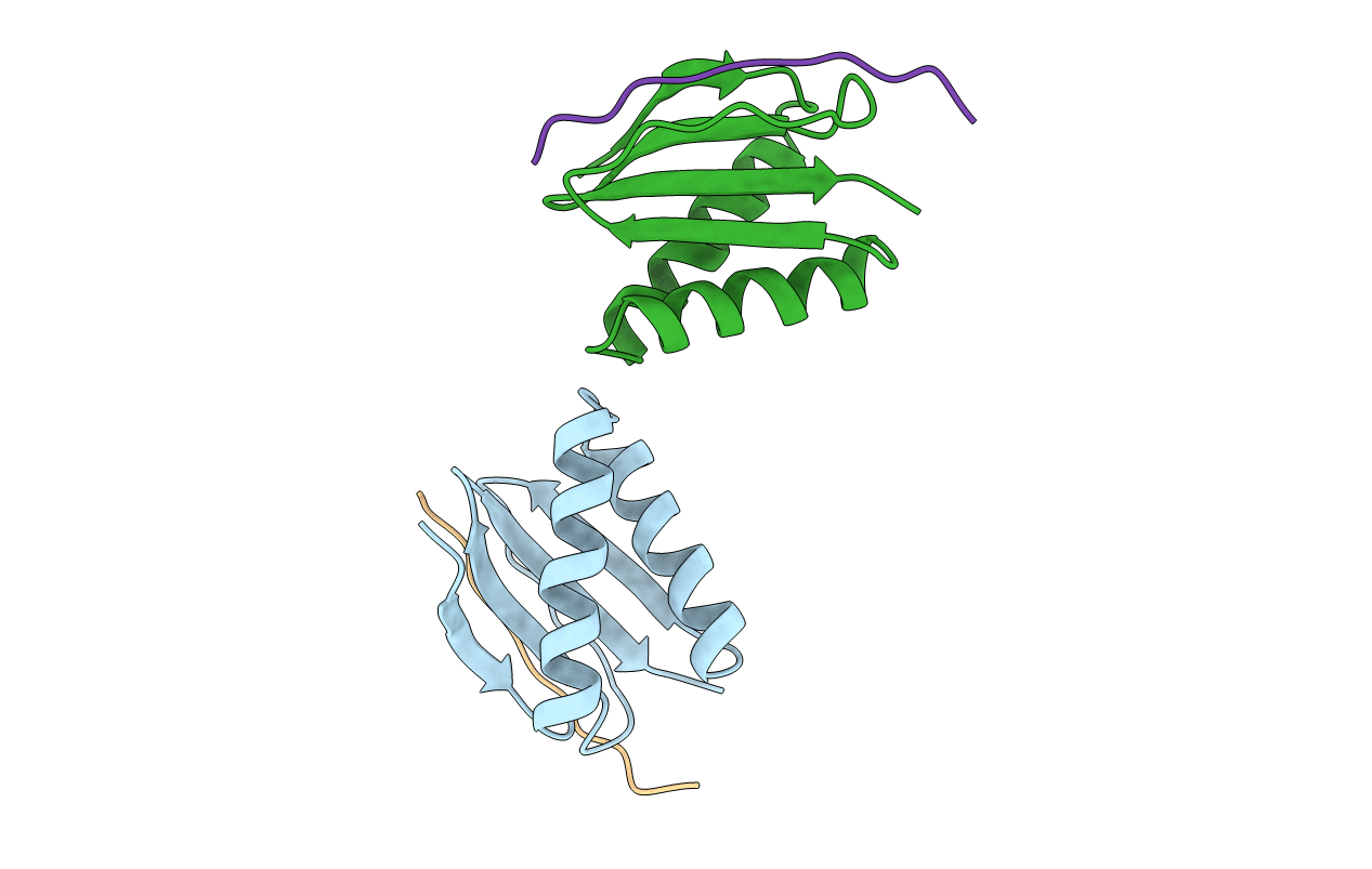
STRUCTURE OF THE HUMAN PIN/LC8 DIMER WITH A BOUND PEPTIDE
Biological Source:
Source Organism:
Homo sapiens (Taxon ID: 9606)
Mus musculus (Taxon ID: 10090)
Host Organism:
Method Details:
Experimental Method:
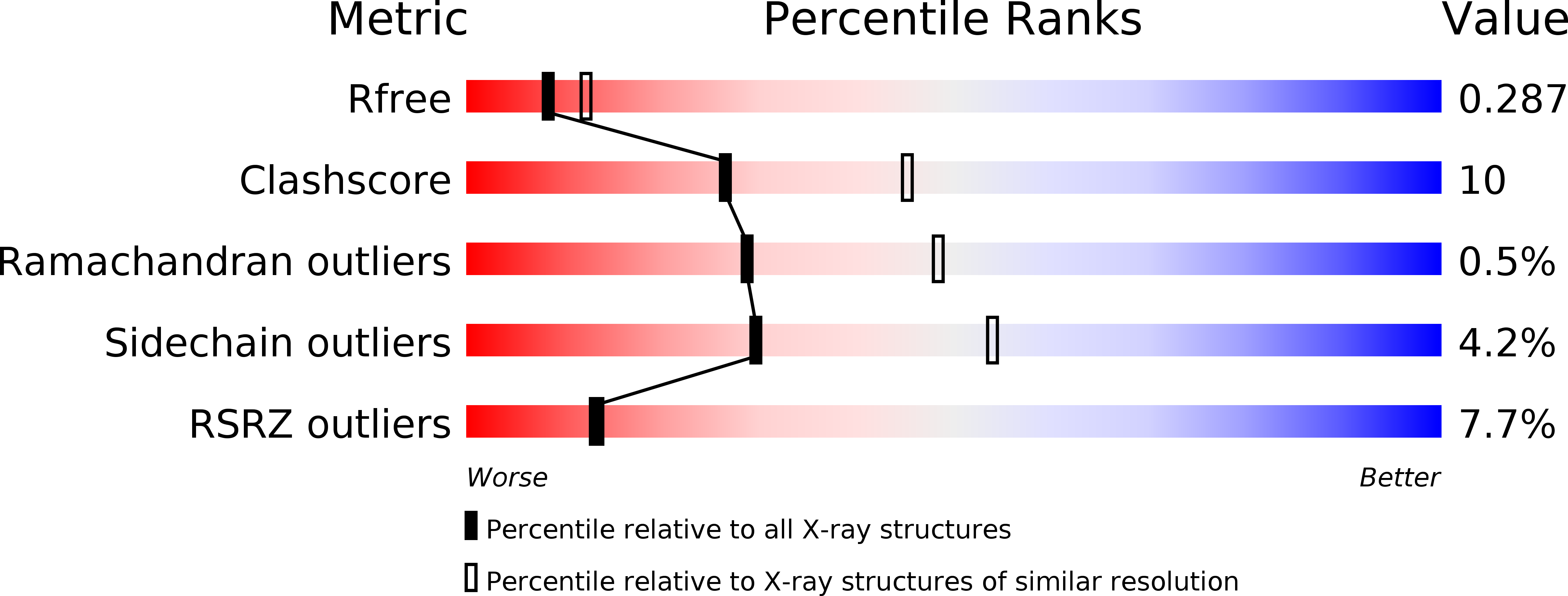
Resolution:
2.50 Å
R-Value Free:
0.28
R-Value Work:
0.24
Space Group:
I 41 2 2
Macromolecular Entities
Structures with similar UniProt ID
Protein Blast
Polymer Type:polypeptide(L)
Molecule:Dynein light chain 1, cytoplasmic
Gene (Uniprot):DYNLL1
Chain IDs:A, B
Chain Length:85
Number of Molecules:2
Biological Source:Homo sapiens
Structures with similar UniProt ID
Protein Blast
Polymer Type:polypeptide(L)
Molecule:Nitric oxide synthase 1
Chain IDs:C, D
Chain Length:13
Number of Molecules:2
Biological Source:Mus musculus
Primary Citation
Structure of the PIN/LC8 dimer with a bound peptide.
Nat.Struct.Biol. 6 735 740 (1999)
PMID: 10426949 DOI: 10.1038/11501

Abstact

The structure of the protein known both as neuronal nitric oxide synthase inhibitory protein, PIN (protein inhibitor of nNOS), and also as the 8 kDa dynein light chain (LC8) has been solved by X-ray diffraction. Two PIN/LC8 monomers related by a two-fold axis form a rectangular dimer. Two pairs of alpha-helices cover opposite faces, and each pair of helices packs against a beta-sheet with five antiparallel beta-strands. Each five-stranded beta-sheet contains four strands from one monomer and a fifth strand from the other monomer. A 13-residue peptide from nNOS is bound to the dimer in a deep hydrophobic groove as a sixth antiparallel beta-strand. The structure provides key insights into dimerization of and peptide binding by the multifunctional PIN/LC8 protein.

Legend

Protein

Chemical

Disease

Primary Citation of related structures
Feedback Form
Name
Email
Institute
Feedback