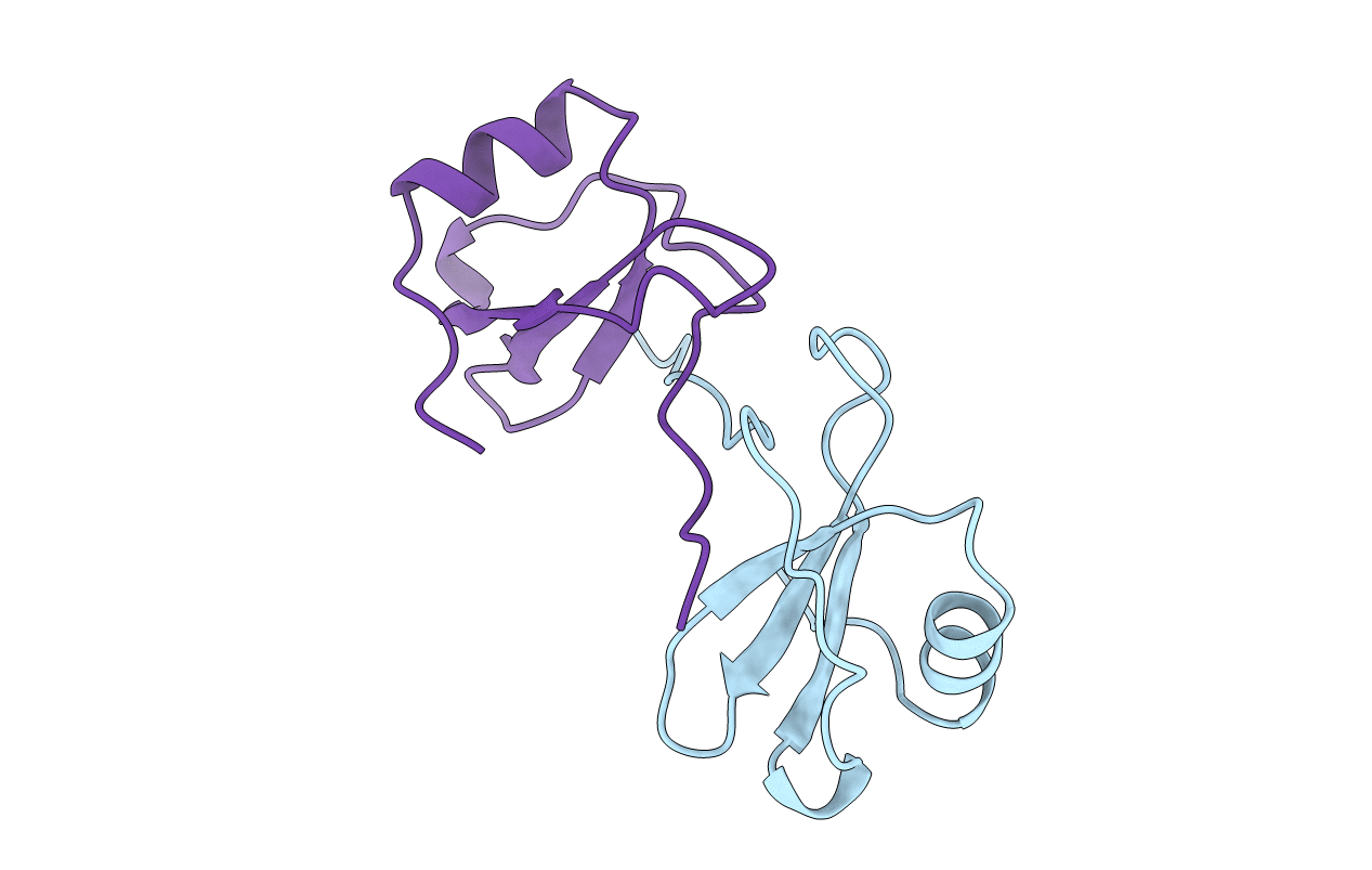
1CM9 image
Entry Detail
PDB ID:
1CM9
Keywords:
Title:
CRYSTAL STRUCTURE OF VIRAL MACROPHAGE INFLAMMATORY PROTEIN-II
Biological Source:
Source Organism:
PDB Version:
Deposition Date:
1999-05-19
Release Date:
1999-06-24
Method Details:
Experimental Method:
Resolution:
2.10 Å
R-Value Free:
0.27
R-Value Work:
0.24
R-Value Observed:
0.24
Space Group:
C 1 2 1
Macromolecular Entities
Polymer Type:polypeptide(L)
Description:PROTEIN (VIRAL MACROPHAGE INFLAMMATORY PROTEIN-II)
Chain IDs:A, B
Chain Length:74
Number of Molecules:2
Biological Source:Human herpesvirus 8
Modified Residue
Compound ID Chain ID Parent Comp ID Details 2D Image
MSE A MET SELENOMETHIONINE
Primary Citation
Comparison of the structure of vMIP-II with eotaxin-1, RANTES, and MCP-3 suggests a unique mechanism for CCR3 activation.
Biochemistry 39 12837 12844 (2000)
PMID: 11041848 DOI: 10.1021/bi001166f

Abstact

Herpesvirus-8 macrophage inflammatory protein-II (vMIP-II) binds a uniquely wide spectrum of chemokine receptors. We report the X-ray structure of vMIP-II determined to 2.1 A resolution. Like RANTES, vMIP-II crystallizes as a dimer and displays the conventional chemokine tertiary fold. We have compared the surface topology and electrostatic potential of vMIP-II to those of eotaxin-1, RANTES, and MCP-3, three CCR3 physiological agonists with known three-dimensional structures. Surface epitopes identified on RANTES to be involved in binding to CCR3 are mimicked on the eotaxin-1 and MCP-3 surface. However, the surface topology of vMIP-II in these regions is markedly different. The results presented here indicate that the structural basis for interaction with the chemokine receptor CCR3 by vMIP-II is different from that for the physiological agonists eotaxin-1, RANTES, and MCP-3. These differences on vMIP-II may be a consequence of its broad-range receptor recognition capabilities.

Legend

Protein

Chemical

Disease

Primary Citation of related structures