1CL2 image
Deposition Date 1997-09-04
Release Date 1998-09-09
Last Version Date 2024-02-07
Entry Detail
PDB ID:
1CL2
Title:
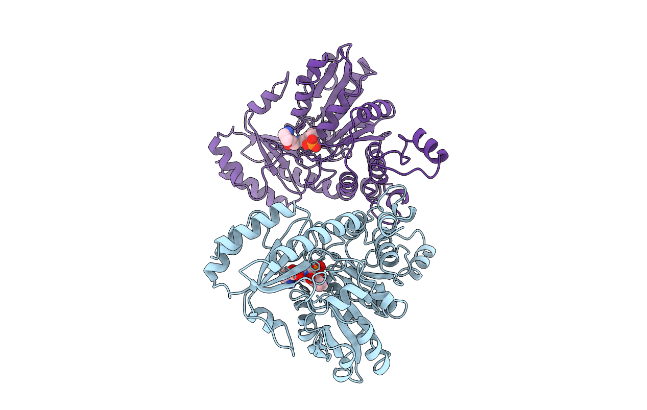
CYSTATHIONINE BETA-LYASE (CBL) FROM ESCHERICHIA COLI IN COMPLEX WITH AMINOETHOXYVINYLGLYCINE
Biological Source:
Source Organism:
Host Organism:
Method Details:
Experimental Method:
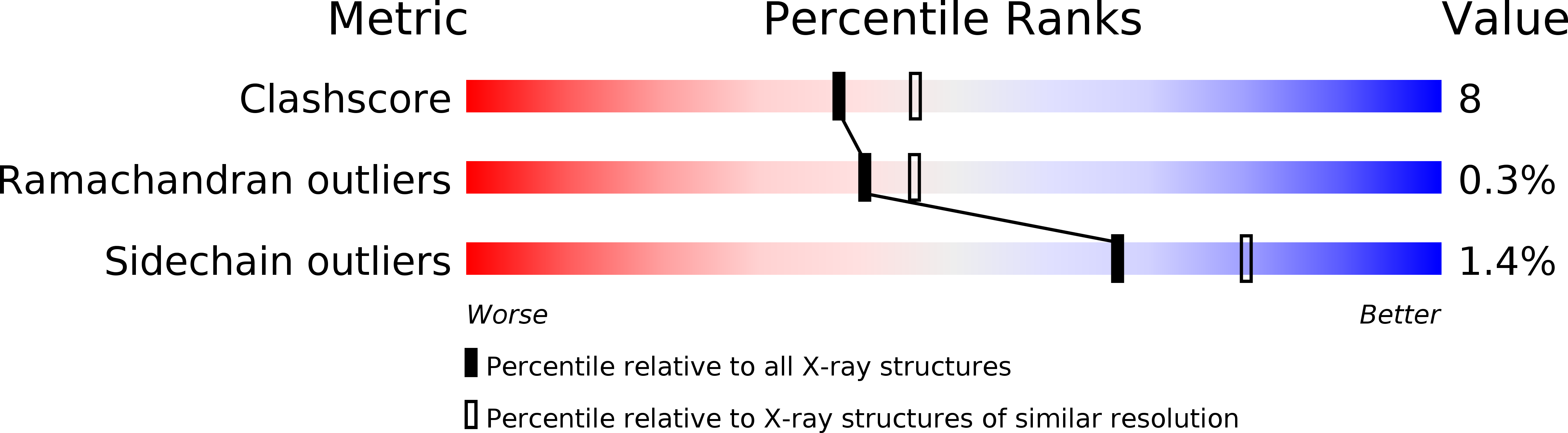
Resolution:
2.20 Å
R-Value Work:
0.16
R-Value Observed:
0.16
Space Group:
C 2 2 21
Macromolecular Entities
Polymer Type:polypeptide(L)
Molecule:CYSTATHIONINE BETA-LYASE
Gene (Uniprot):metC
Chain IDs:A, B
Chain Length:395
Number of Molecules:2
Biological Source:Escherichia coli
Primary Citation
Slow-binding inhibition of Escherichia coli cystathionine beta-lyase by L-aminoethoxyvinylglycine: a kinetic and X-ray study.
Biochemistry 36 12633 12643 (1997)
PMID: 9376370 DOI: 10.1021/bi970630m

Abstact

The pyridoxal 5'-phosphate (PLP)-dependent cystathionine beta-lyase (CBL) was previously found to be inhibited by the natural toxins rhizobitoxine and l-aminoethoxyvinylglycine (AVG). The present study characterizes the interaction of Escherichia coli CBL with AVG and methoxyvinylglycine (MVG) by a combination of kinetic methods and X-ray crystallography. Upon AVG treatment, time-dependent, slow-binding inhibition [Morrison, J. F. (1982) Trends Biochem. Sci. 7, 102-105] was observed due to the generation of a long-lived, slowly dissociating enzyme-inhibitor complex. Kinetic analysis revealed a one-step inhibition mechanism (CBL + AVG --> CBLAVG, Ki = 1.1 +/- 0.3 microM) with an association rate constant (k1) of 336 +/- 40 M-1 s-1. This value is several orders of magnitude lower than typical bimolecular rate constants of ES formation, suggesting that additional steps occur before formation of the first detectable CBLAVG complex. Loss of activity is paralleled by the conversion of the pyridoxaldimine 426 nm chromophore to a 341 nm-absorbing species. On the basis of the recently solved structure of native CBL [Clausen, T., et al. (1996) J. Mol. Biol. 262, 202-224], it was possible to elucidate the X-ray structure of the CBLAVG complex and to refine it to an R-factor of 16.4% at 2.2 A resolution. The refined structure reveals the geometry of the bound inhibitor and its interactions with residues in the active site of CBL. Both the X-ray structure and the absorbance spectrum of the CBLAVG complex are compatible with a ketimine as the reaction product. Thus, the inhibitor seems to bind in a similar way to CBL as the substrate, but after alpha-proton abstraction, the reaction proceeds in a CBL nontypical manner, i.e. protonation of PLP-C4', resulting in the "dead-end" ketimine PLP derivative. The CBLAVG structure furthermore suggests a binding mode for rhizobitoxine and explains the failure of MVG to inhibit CBL.

Legend

Protein

Chemical

Disease

Primary Citation of related structures