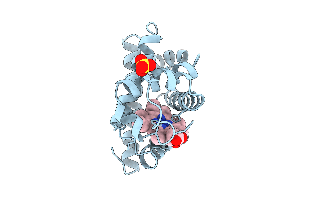
1CIO image
Entry Detail
PDB ID:
1CIO
Title:
RECOMBINANT SPERM WHALE MYOGLOBIN I99V MUTANT (MET)
Biological Source:
Source Organism:
Host Organism:
PDB Version:
Deposition Date:
1999-04-01
Release Date:
1999-04-09
Method Details:
Experimental Method:
Resolution:
1.60 Å
R-Value Free:
0.18
R-Value Observed:
0.14
Space Group:
P 6
Macromolecular Entities
Polymer Type:polypeptide(L)
Description:PROTEIN (MYOGLOBIN)
Mutations:INITIATOR MET, I99V, D122N
Chain IDs:A
Chain Length:154
Number of Molecules:1
Biological Source:Physeter catodon
Primary Citation
Waterproofing the heme pocket. Role of proximal amino acid side chains in preventing hemin loss from myoglobin.
J.Biol.Chem. 276 9093 9100 (2001)
PMID: 11084036 DOI: 10.1074/jbc.M008593200

Abstact

The ability of myoglobin to bind oxygen reversibly depends critically on retention of the heme prosthetic group. Globin side chains at the Leu(89)(F4), His(97)(FG3), Ile(99)(FG5), and Leu(104)(G5) positions on the proximal side of the heme pocket strongly influence heme affinity. The roles of these amino acids in preventing heme loss have been examined by determining high resolution structures of 14 different mutants at these positions using x-ray crystallography. Leu(89) and His(97) are important surface amino acids that interact either sterically or electrostatically with the edges of the porphyrin ring. Ile(99) and Leu(104) are located in the interior region of the proximal pocket beneath ring C of the heme prosthetic group. The apolar amino acids Leu(89), Ile(99), and Leu(104) "waterproof" the heme pocket by forming a barrier to solvent penetration, minimizing the size of the proximal cavity, and maintaining a hydrophobic environment. Substitutions with smaller or polar side chains at these positions result in exposure of the heme to solvent, the appearance of crystallographically defined water molecules in or near the proximal pocket, and large increases in the rate of hemin loss. Thus, the naturally occurring amino acid side chains at these positions serve to prevent hydration of the His(93)-Fe(III) bond and are highly conserved in all known myoglobins and hemoglobins.

Legend

Protein

Chemical

Disease

Primary Citation of related structures