1CIM image
Entry Detail
PDB ID:
1CIM
Keywords:
Title:
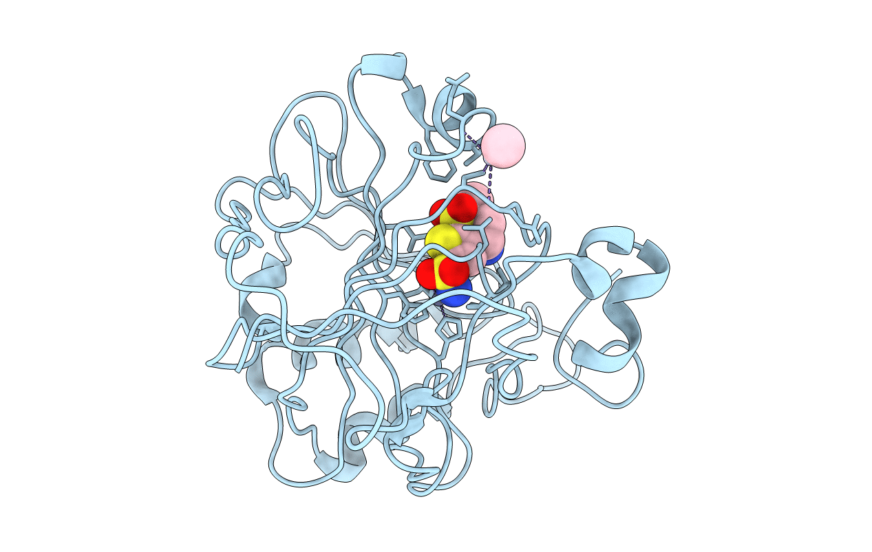
THE POSITIONS OF HIS-64 AND A BOUND WATER IN HUMAN CARBONIC ANHYDRASE II UPON BINDING THREE STRUCTURALLY RELATED INHIBITORS
Biological Source:
Source Organism:
PDB Version:
Deposition Date:
1993-10-20
Release Date:
1994-01-31
Method Details:
Experimental Method:
Resolution:
2.10 Å
Space Group:
P 1 21 1
Macromolecular Entities
Polymer Type:polypeptide(L)
Description:CARBONIC ANHYDRASE II
Chain IDs:A
Chain Length:259
Number of Molecules:1
Biological Source:Homo sapiens
Primary Citation
Positions of His-64 and a bound water in human carbonic anhydrase II upon binding three structurally related inhibitors.
Protein Sci. 3 118 125 (1994)
PMID: 8142888

Abstact

The 3-dimensional structure of human carbonic anhydrase II (HCAII; EC 4.2.1.1) complexed with 3 structurally related inhibitors, 1a, 1b, and 1c, has been determined by X-ray crystallographic methods. The 3 inhibitors (1a = C8H12N2O4S3) vary only in the length of the substituent on the 4-amino group: 1a, proton; 1b, methyl; and 1c, ethyl. The binding constants (Ki's) for 1a, 1b, and 1c to HCAII are 1.52, 1.88, and 0.37 nM, respectively. These structures were solved to learn if any structural cause could be found for the difference in binding. In the complex with inhibitors 1a and 1b, electron density can be observed for His-64 and a bound water molecule in the native positions. When inhibitor 1c is bound, the side chain attached to the 4-amino group is positioned so that His-64 can only occupy the alternate position and the bound water is absent. While a variety of factors contribute to the observed binding constants, the major reason 1c binds tighter to HCAII than does 1a or 1b appears to be entropy: the increase in entropy when the bound water molecule is released contributes to the increase in binding and overcomes the small penalty for putting the His-64 side chain in a higher energy state.

Legend

Protein

Chemical

Disease

Primary Citation of related structures