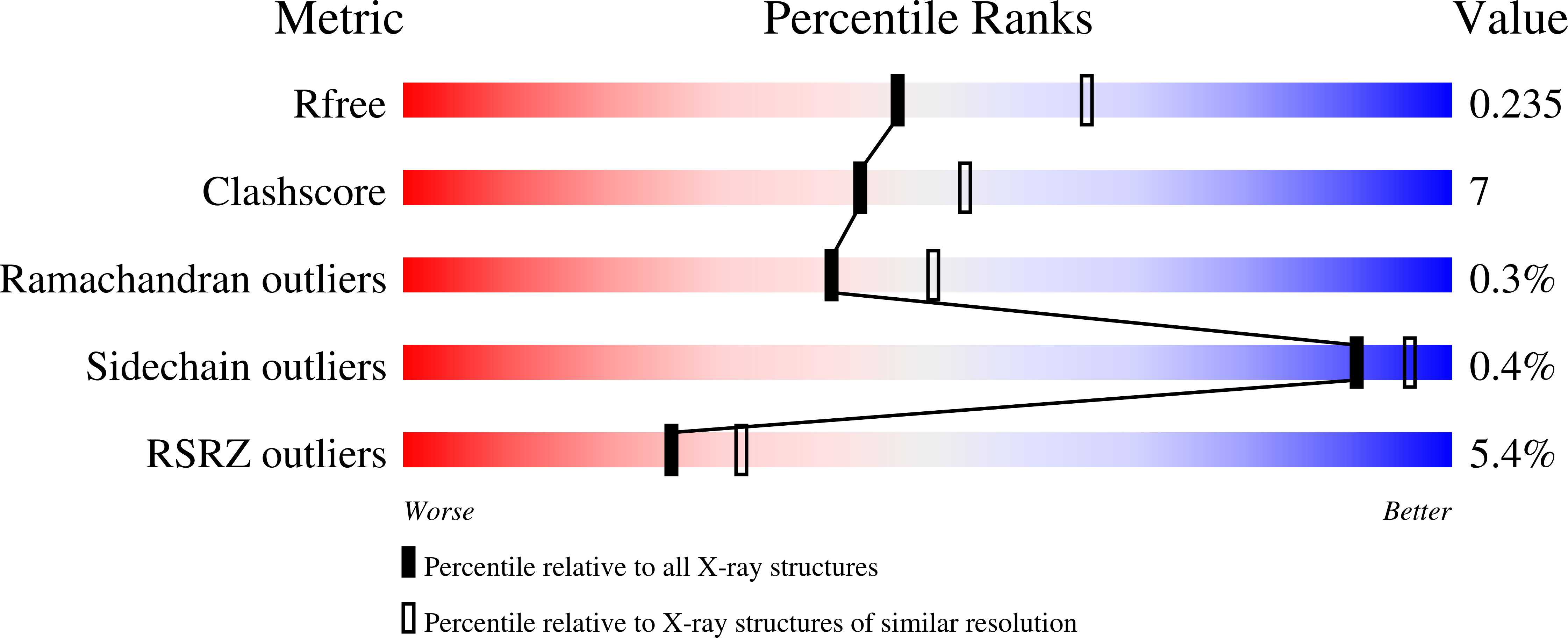
1CH0 image
Deposition Date 1999-03-30
Release Date 1999-12-22
Last Version Date 2024-11-13
Entry Detail
PDB ID:
1CH0
Keywords:
Title:
RNASE T1 VARIANT WITH ALTERED GUANINE BINDING SEGMENT
Biological Source:
Source Organism:
Host Organism:
Method Details:
Experimental Method:
Resolution:
2.30 Å
R-Value Free:
0.24
R-Value Work:
0.19
Space Group:
P 21 21 21
Macromolecular Entities
Polymer Type:polypeptide(L)
Molecule:PROTEIN (RIBONUCLEASE T1)
Gene (Uniprot):rntA
Mutations:Q25K, K41E, Y42F, N43R, Y45W, E46Q
Chain IDs:A, B, C
Chain Length:104
Number of Molecules:3
Biological Source:Aspergillus oryzae
Primary Citation
Structural analysis of an RNase T1 variant with an altered guanine binding segment.
J.Mol.Biol. 294 1231 1238 (1999)
PMID: 10600381 DOI: 10.1006/jmbi.1999.3324

Abstact

The ribonuclease T1 variant 9/5 with a guanine recognition segment, altered from the wild-type amino acid sequence 41-KYNNYE-46 to 41-EFRNWQ-46, has been cocrystallised with the specific inhibitor 2'-GMP. The crystal structure has been refined to a crystallographic R factor of 0.198 at 2.3 A resolution. Despite a size reduction of the binding pocket, pushing the inhibitor outside by 1 A, 2'-GMP is fixed to the primary recognition site due to increased aromatic stacking interactions. The phosphate group of 2'-GMP is located about 4.2 A apart from its position in wild-type ribonuclease T1-2'-GMP complexes, allowing a Ca(2+), coordinating this phosphate group, to enter the binding pocket. The crystallographic data can be aligned with the kinetic characterisation of the variant, showing a reduction of both, guanine affinity and turnover rate. The presence of Ca(2+) was shown to inhibit variant 9/5 and wild-type enzyme to nearly the same extent.

Legend

Protein

Chemical

Disease

Primary Citation of related structures