1CFP image
Deposition Date 1996-06-04
Release Date 1997-03-12
Last Version Date 2024-05-22
Entry Detail
PDB ID:
1CFP
Title:
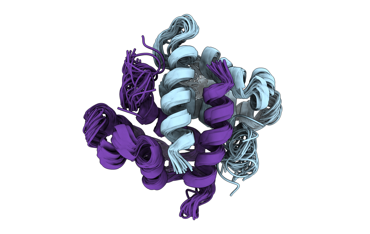
S100B (S100BETA) NMR DATA WAS COLLECTED FROM A SAMPLE OF CALCIUM FREE PROTEIN AT PH 6.3 AND A TEMPERATURE OF 311 K AND 1.7-6.9 MM CONCENTRATION, 25 STRUCTURES
Biological Source:
Source Organism:
Bos taurus (Taxon ID: 9913)
Host Organism:
Method Details:
Experimental Method:
Conformers Calculated:
91
Conformers Submitted:
25
Selection Criteria:
TOTAL ENERGY
Macromolecular Entities
Structures with similar UniProt ID
Protein Blast
Polymer Type:polypeptide(L)
Molecule:S100B
Gene (Uniprot):S100B
Mutagens:N-TERMINAL METHIONINE REPLACES N-TERMINAL ACETYL GROUP OF THE NATURAL PROTEIN
Chain IDs:A, B
Chain Length:92
Number of Molecules:2
Biological Source:Bos taurus
Ligand Molecules
Primary Citation
The solution structure of the bovine S100B protein dimer in the calcium-free state.
Structure 4 1041 1052 (1996)
PMID: 8805590 DOI: 10.1016/S0969-2126(96)00111-6

Abstact

BACKGROUND S100B (S100beta) is a member of the S100 family of small calcium-binding proteins: members of this family contain two helix-loop-helix calcium-binding motifs and interact with a wide range of proteins involved mainly in the cytoskeleton and cell proliferation. S100B is a neurite-extension factor and levels of S100B are elevated in the brains of patients with Alzheimer's disease or Down's syndrome: the pattern of S100B overexpression in Alzheimer's disease correlates with the pattern of neuritic-plaque formation. Identification of a growing class of S100 proteins and the likely neurochemical importance of S100B make the determination of the structure of S100B of interest. RESULTS We have used NMR to determine the structure of the reduced S100B homodimer in the absence of calcium. Each monomer consists of a four-helix bundle, arranged in the dimer in an antiparallel fashion. The fourth helix of each monomer runs close to the equivalent helix of the other monomer for almost its full length, extending the hydrophobic core through the interface. The N-terminal, but not the C-terminal, calcium-binding loop is similar to the equivalent loop in the monomeric S100 protein calbindin and is in a conformation ready to bind calcium. CONCLUSIONS The novel dimer structure reported previously for calcyclin (S100A6) is the common fold for the dimeric S100B proteins. Calcium binding to the C-terminal calcium-binding loop would be expected to require a conformational change, which might provide a signal for activation. The structure suggests regions of the molecule likely to be involved in interactions with effector molecules.

Legend

Protein

Chemical

Disease

Primary Citation of related structures