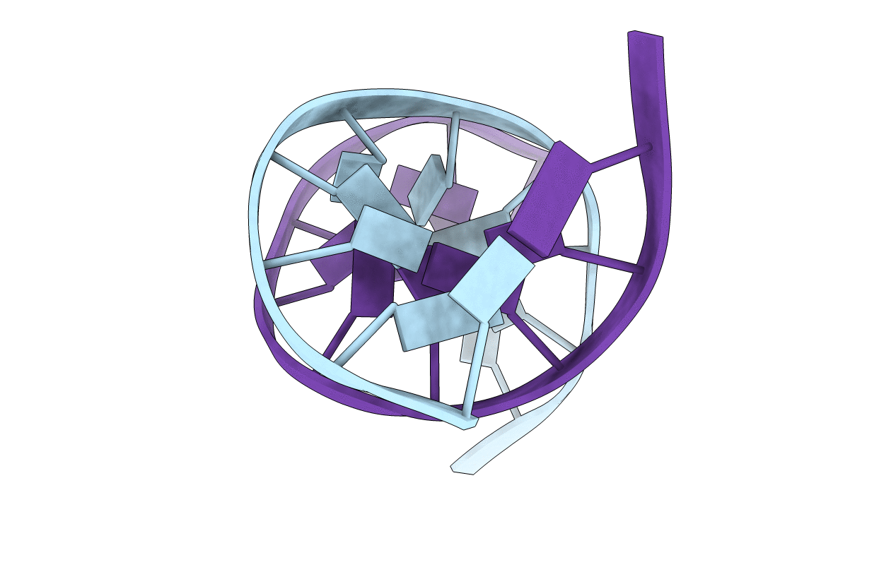
1CFL image
Deposition Date 1999-03-19
Release Date 1999-05-28
Last Version Date 2023-12-27
Entry Detail
PDB ID:
1CFL
Keywords:
Title:
DNA DECAMER DUPLEX CONTAINING T5-T6 PHOTOADDUCT
Biological Source:
Source Organism:
Method Details:
Experimental Method:
Conformers Submitted:
1
Selection Criteria:
LEAST RESTRAINT VIOLATION
Macromolecular Entities
Polymer Type:polydeoxyribonucleotide
Molecule:DNA (5'-D(*CP*GP*CP*AP*(64T)P*TP*AP*CP*GP*C)-3')
Chain IDs:A
Chain Length:10
Number of Molecules:1
Biological Source:
Polymer Type:polydeoxyribonucleotide
Molecule:DNA (5'-D(*GP*CP*GP*TP*GP*AP*TP*GP*CP*G)-3')
Chain IDs:B
Chain Length:10
Number of Molecules:1
Biological Source:
Modified Residue
Compound ID Chain ID Parent Comp ID Details 2D Image
64T A DT 5-HYDROXY-THYMIDINE-5'-MONOPHOSPHATE
Ligand Molecules
Primary Citation
Solution structure of a DNA decamer duplex containing the stable 3' T.G base pair of the pyrimidine(6-4)pyrimidone photoproduct [(6-4) adduct]: implications for the highly specific 3' T --> C transition of the (6-4) adduct.
Proc.Natl.Acad.Sci.USA 96 6632 6636 (1999)
PMID: 10359763 DOI: 10.1073/pnas.96.12.6632

Abstact

The pyrimidine(6-4)pyrimidone photoproduct [(6-4) adduct] is one of the major photoproducts induced by UV irradiation of DNA and occurs at TpT sites. The (6-4) adduct is highly mutagenic and leads most often to a 3' T --> C transition with 85% replicating error frequency [LeClerc, J. E., Borden, A. & Lawrence, C. W. (1991) Proc. Natl. Acad. Sci. USA 88, 9685-9689]. To determine the origin of the specific 3' T --> C transition of the (6-4) adduct, we have used experimental NMR restraints and molecular dynamics to determine the solution structure of a (6-4)-lesion DNA decamer duplex that contains a mismatched base pair between the 3' T residue and an opposed G residue. Normal Watson-Crick-type hydrogen bonding is retained at the 5' T of the lesion site. The O2 carbonyl of the 3' T residue forms hydrogen bonds with the imino and amino protons of the opposed G residue. This potential hydrogen bonding stabilizes the overall helix and restores the highly distorted conformation of the (6-4) adduct to the typical B-form-like DNA structure. This structural feature can explain the marked preference for the insertion of an A residue opposite the 5' T and a G residue opposite the 3' T of the (6-4) lesion during trans-lesion synthesis. Thus these insertions yield the predominant 3' T --> C transition.

Legend

Protein

Chemical

Disease

Primary Citation of related structures