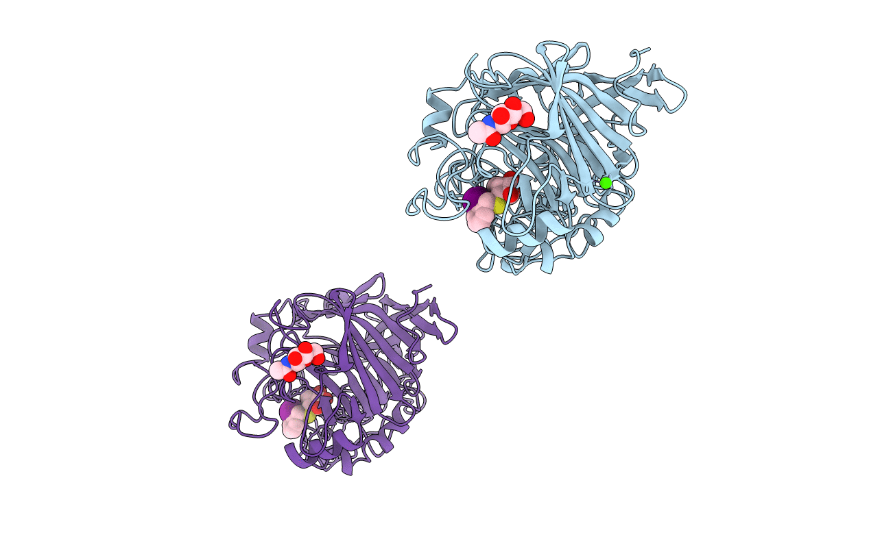
1CEL image
Deposition Date 1994-05-17
Release Date 1994-11-01
Last Version Date 2024-10-30
Entry Detail
PDB ID:
1CEL
Title:
THE THREE-DIMENSIONAL CRYSTAL STRUCTURE OF THE CATALYTIC CORE OF CELLOBIOHYDROLASE I FROM TRICHODERMA REESEI
Biological Source:
Source Organism:
Method Details:
Experimental Method:
Resolution:
1.80 Å
R-Value Work:
0.18
R-Value Observed:
0.18
Space Group:
P 21 21 2
Macromolecular Entities
Polymer Type:polypeptide(L)
Molecule:1,4-BETA-D-GLUCAN CELLOBIOHYDROLASE I
Chain IDs:A, B
Chain Length:434
Number of Molecules:2
Biological Source:Trichoderma reesei
Modified Residue
Compound ID Chain ID Parent Comp ID Details 2D Image
ASN A ASN GLYCOSYLATION SITE
PCA A GLN PYROGLUTAMIC ACID
Primary Citation
The three-dimensional crystal structure of the catalytic core of cellobiohydrolase I from Trichoderma reesei.
Science 265 524 528 (1994)
PMID: 8036495

Abstact

Cellulose is the major polysaccharide of plants where it plays a predominantly structural role. A variety of highly specialized microorganisms have evolved to produce enzymes that either synergistically or in complexes can carry out the complete hydrolysis of cellulose. The structure of the major cellobiohydrolase, CBHI, of the potent cellulolytic fungus Trichoderma reesei has been determined and refined to 1.8 angstrom resolution. The molecule contains a 40 angstrom long active site tunnel that may account for many of the previously poorly understood macroscopic properties of the enzyme and its interaction with solid cellulose. The active site residues were identified by solving the structure of the enzyme complexed with an oligosaccharide, o-iodobenzyl-1-thio-beta-cellobioside. The three-dimensional structure is very similar to a family of bacterial beta-glucanases with the main-chain topology of the plant legume lectins.

Legend

Protein

Chemical

Disease

Primary Citation of related structures