1CEK image
Deposition Date 1999-03-09
Release Date 1999-03-11
Last Version Date 2023-12-27
Entry Detail
PDB ID:
1CEK
Title:
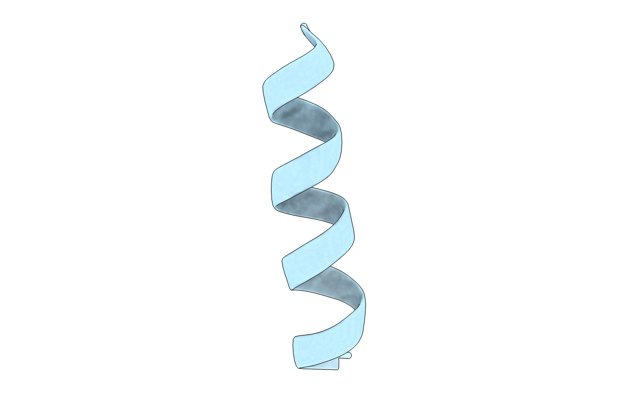
THREE-DIMENSIONAL STRUCTURE OF THE MEMBRANE-EMBEDDED M2 CHANNEL-LINING SEGMENT FROM THE NICOTINIC ACETYLCHOLINE RECEPTOR BY SOLID-STATE NMR SPECTROSCOPY
Biological Source:
Source Organism:
Host Organism:
Method Details:
Experimental Method:
Conformers Calculated:
24
Conformers Submitted:
1
Selection Criteria:
LOWEST ENERGY
Macromolecular Entities
Polymer Type:polypeptide(L)
Molecule:PROTEIN (ACETYLCHOLINE RECEPTOR M2)
Gene (Uniprot):Chrnd
Chain IDs:A
Chain Length:25
Number of Molecules:1
Biological Source:Rattus norvegicus
Ligand Molecules
Primary Citation
Structures of the M2 channel-lining segments from nicotinic acetylcholine and NMDA receptors by NMR spectroscopy.
Nat.Struct.Biol. 6 374 379 (1999)
PMID: 10201407 DOI: 10.1038/7610

Abstact

The structures of functional peptides corresponding to the predicted channel-lining M2 segments of the nicotinic acetylcholine receptor (AChR) and of a glutamate receptor of the NMDA subtype (NMDAR) were determined using solution NMR experiments on micelle samples, and solid-state NMR experiments on bilayer samples. Both M2 segments form straight transmembrane alpha-helices with no kinks. The AChR M2 peptide inserts in the lipid bilayer at an angle of 12 degrees relative to the bilayer normal, with a rotation about the helix long axis such that the polar residues face the N-terminal side of the membrane, which is assigned to be intracellular. A model built from these solid-state NMR data, and assuming a symmetric pentameric arrangement of M2 helices, results in a funnel-like architecture for the channel, with the wide opening on the N-terminal intracellular side.

Legend

Protein

Chemical

Disease

Primary Citation of related structures