Deposition Date
1995-09-29
Release Date
1996-03-08
Last Version Date
2024-02-07
Entry Detail
PDB ID:
1CDO
Keywords:
Title:
ALCOHOL DEHYDROGENASE (E.C.1.1.1.1) (EE ISOZYME) COMPLEXED WITH NICOTINAMIDE ADENINE DINUCLEOTIDE (NAD), AND ZINC
Biological Source:
Source Organism(s):
Gadus callarias (Taxon ID: 8053)
Method Details:
Experimental Method:
Resolution:
2.05 Å
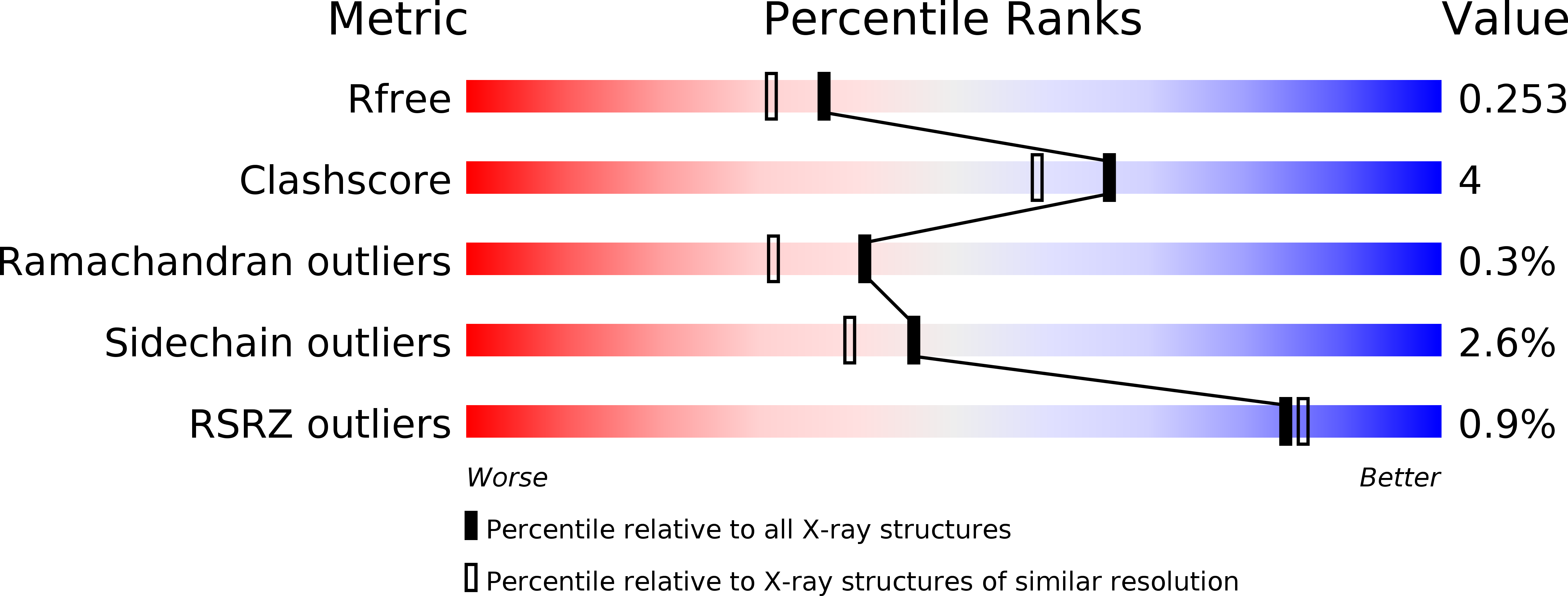
R-Value Free:
0.24
R-Value Work:
0.17
R-Value Observed:
0.17
Space Group:
P 1 21 1