Deposition Date
1984-08-10
Release Date
1984-10-29
Last Version Date
2024-11-13
Entry Detail
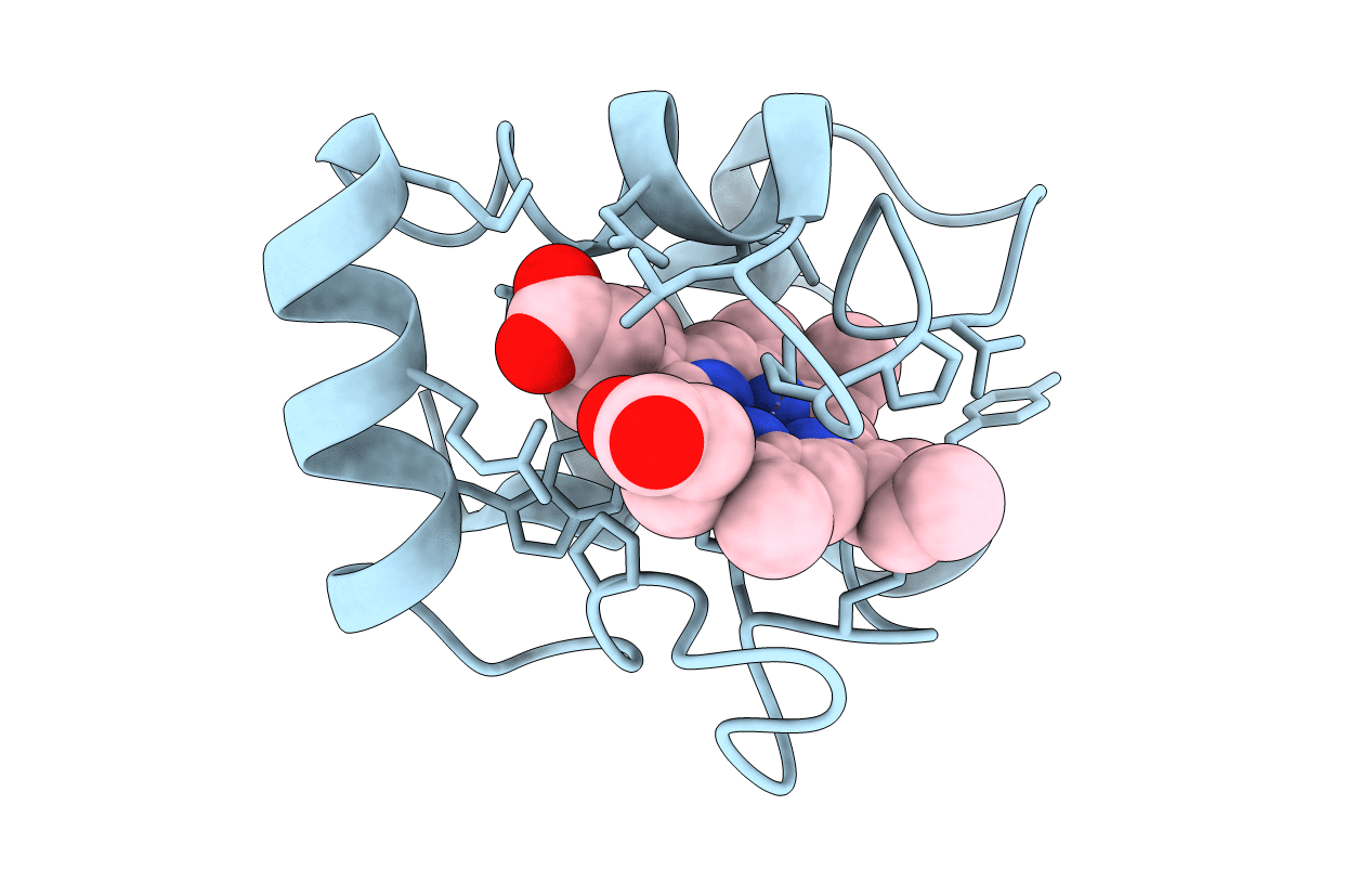
PDB ID:
1CC5
Keywords:
Title:
CRYSTAL STRUCTURE OF AZOTOBACTER CYTOCHROME C5 AT 2.5 ANGSTROMS RESOLUTION
Biological Source:
Source Organism(s):
Azotobacter vinelandii (Taxon ID: 354)
Method Details: