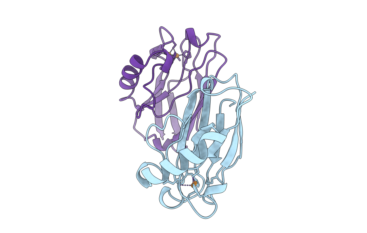
1CC3 image
Deposition Date 1999-03-03
Release Date 1999-12-23
Last Version Date 2024-11-13
Entry Detail
PDB ID:
1CC3
Title:
PURPLE CUA CENTER
Biological Source:
Source Organism:
Host Organism:
Method Details:
Experimental Method:
Resolution:
1.65 Å
R-Value Free:
0.26
R-Value Work:
0.20
R-Value Observed:
0.20
Space Group:
P 1 21 1
Macromolecular Entities
Polymer Type:polypeptide(L)
Molecule:PROTEIN (CUA AZURIN)
Gene (Uniprot):azu
Chain IDs:A, B
Chain Length:130
Number of Molecules:2
Biological Source:Pseudomonas aeruginosa
Primary Citation
Structural basis of electron transfer modulation in the purple CuA center.
Biochemistry 38 5677 5683 (1999)
PMID: 10231517 DOI: 10.1021/bi9901634

Abstact

The X-ray structure of an engineered purple CuA center in azurin from Pseudomonas aeruginosa has been determined and refined at 1.65 A resolution. Two independent purple CuA azurin molecules are in the asymmetric unit of a new P21 crystal, and they have nearly identical conformations (rmsd of 0.27 A for backbone atoms). The purple CuA azurin was produced by the loop-engineering strategy, and the resulting overall structure is unperturbed. The insertion of a slightly larger Cu-binding loop into azurin causes the two structural domains of azurin to move away from each other. The high-resolution structure reveals the detailed environment of the delocalized mixed-valence [Cu(1.5).Cu(1.5)] binuclear purple CuA center, which serves as a useful reference model for other native proteins, and provides a firm basis for understanding results from spectroscopic and functional studies of this class of copper center in biology. The two independent Cu-Cu distances of 2.42 and 2.35 A (with respective concomitant adjustments of ligand-Cu distances) are consistent with that (2.39 A) obtained from X-ray absorption spectroscopy with the same molecule, and are among the shortest Cu-Cu bonds observed to date in proteins or inorganic complexes. A comparison of the purple CuA azurin structure with those of other CuA centers reveals an important relationship between the angular position of the two His imidazole rings with respect to the Cu2S2(Cys) core plane and the distance between the Cu and the axial ligand. This relationship strongly suggests that the fine structural variation of different CuA centers can be correlated with the angular positions of the two histidine rings because, from these positions, one can predict the relative axial ligand interactions, which are responsible for modulating the Cu-Cu distance and the electron transfer properties of the CuA centers.

Legend

Protein

Chemical

Disease

Primary Citation of related structures