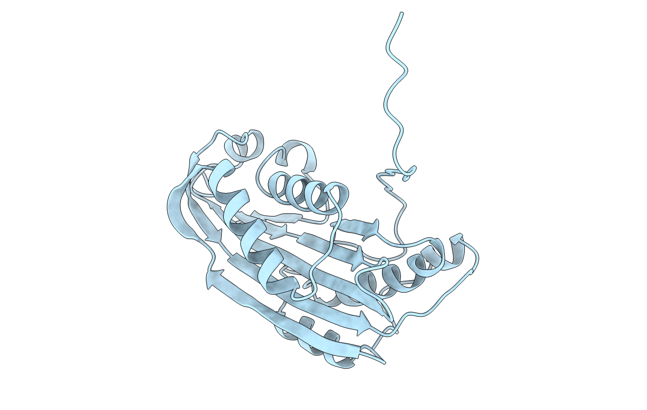
1CBY image
Deposition Date 1995-09-05
Release Date 1996-10-14
Last Version Date 2024-02-07
Entry Detail
PDB ID:
1CBY
Keywords:
Title:
DELTA-ENDOTOXIN
Biological Source:
Host Organism:
Method Details:
Experimental Method:
Resolution:
2.60 Å
R-Value Free:
0.26
R-Value Work:
0.19
R-Value Observed:
0.19
Space Group:
P 61 2 2
Macromolecular Entities
Structures with similar UniProt ID
Protein Blast
Polymer Type:polypeptide(L)
Molecule:DELTA-ENDOTOXIN CYTB
Gene (Uniprot):cyt2Aa1
Chain IDs:A
Chain Length:259
Number of Molecules:1
Biological Source:Bacillus thuringiensis serovar kyushuensis
Primary Citation
Structure of the mosquitocidal delta-endotoxin CytB from Bacillus thuringiensis sp. kyushuensis and implications for membrane pore formation.
J.Mol.Biol. 257 129 152 (1996)
PMID: 8632451 DOI: 10.1006/jmbi.1996.0152

Abstact

The delta-endotoxin CytB, found in parasporal inclusions of Bacillus thuringiensis subspecies kyushuensis, is a membrane pore-forming protein which is lethal to the larvae of Dipteran insects and broadly cytolytic in vitro. The crystal structure of CytB in the protoxin form has been determined by isomorphous replacement using heavy-atom derivatives of both the wild-type protein and an engineered cysteine mutant. The atomic model comprising residues 19 to 245 and 28 bound water molecules has been refined at 2.6 angstrom resolution to a crystallographic R-factor of 19.7% and a free R-factor of 26.1%. CytB has a single domain of alpha/beta architecture but a novel connectivity comprising two outer layers of alpha-helix hairpins wrapped around a mixed beta-sheet. In the protoxin form, CytB is a dimer linked by the intertwined N-terminal strands in a continuous, 12-stranded beta-sheet. Proteolytic processing cleaves the intertwined beta-strands to release the active CytB as a monomer, as well as removing the C-terminal tail to uncover the three-layered core. The homologous toxin CytA should show the same fold. Mutations in CytA that inhibit expression map to the dimer contacts and to the tip of helix pair A-B in contact with the sheet, apparently preventing correct folding. Mutations that inhibit toxicity map to the edge of the beta-sheet adjoining the helix pair C-D and to the sheet face, while mutations on the helix surfaces have no effect. Therefore segments forming the sheet, rather than the amphiphilic but short helices, are responsible for membrane binding and pore formation. A conformational change is postulated by which the helix pair C-D peels away from the sheet to lie on the membrane surface, while the sheet region rearranges to form an oligomeric trans-membrane pore.

Legend

Protein

Chemical

Disease

Primary Citation of related structures