1CBX image
Deposition Date 1991-10-31
Release Date 1994-01-31
Last Version Date 2024-10-30
Entry Detail
PDB ID:
1CBX
Title:
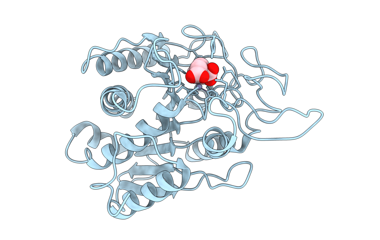
CRYSTAL STRUCTURE OF THE COMPLEX BETWEEN CARBOXYPEPTIDASE A AND THE BIPRODUCT ANALOG INHIBITOR L-BENZYLSUCCINATE AT 2.0 ANGSTROMS RESOLUTION
Biological Source:
Source Organism:
Bos taurus (Taxon ID: 9913)
Method Details:
Experimental Method:
Resolution:
2.00 Å
R-Value Observed:
0.16
Space Group:
P 1 21 1
Macromolecular Entities
Polymer Type:polypeptide(L)
Molecule:CARBOXYPEPTIDASE A
Gene (Uniprot):CPA1
Chain IDs:A
Chain Length:307
Number of Molecules:1
Biological Source:Bos taurus
Primary Citation
Crystal structure of the complex between carboxypeptidase A and the biproduct analog inhibitor L-benzylsuccinate at 2.0 A resolution.
J.Mol.Biol. 223 573 578 (1992)
PMID: 1738164 DOI: 10.1016/0022-2836(92)90671-6

Abstact

The X-ray crystal structure of the carboxypeptidase A-L-benzylsuccinate complex has been refined at 2.0 A resolution to a final R-factor of 0.166. One molecule of the inhibitor binds to the enzyme active site. The terminal carboxylate forms a salt link with the guanidinium group of Arg145 and hydrogen bonds with Tyr248 and Asn144. The second carboxylate group binds to the zinc ion in an asymmetric bidentate fashion replacing the water molecule of the native structure. The zinc ion moves 0.5 A from its position in the native structure to accommodate the inhibitor binding. The overall stereochemistry around the zinc can be considered a distorted tetrahedron, although six atoms of the co-ordinated groups lie within 3.0 A from the zinc ion. The key for the strong inhibitory properties of L-benzylsuccinate can be found in its ability both to co-ordinate the zinc and to form a short carboxyl-carboxylate-type hydrogen bond (2.5 A) with Glu270.

Legend

Protein

Chemical

Disease

Primary Citation of related structures