1CBL image
Deposition Date 1993-02-18
Release Date 1994-07-31
Last Version Date 2024-05-22
Entry Detail
PDB ID:
1CBL
Title:
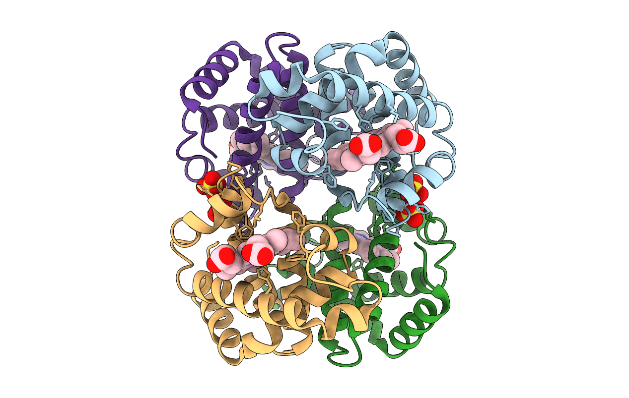
THE 1.9 ANGSTROM STRUCTURE OF DEOXY-BETA4 HEMOGLOBIN: ANALYSIS OF THE PARTITIONING OF QUATERNARY-ASSOCIATED AND LIGAND-INDUCED CHANGES IN TERTIARY STRUCTURE
Biological Source:
Source Organism:
Homo sapiens (Taxon ID: 9606)
Method Details:
Experimental Method:
Resolution:
1.80 Å
R-Value Observed:
0.19
Space Group:
P 1 21 1
Macromolecular Entities
Structures with similar UniProt ID
Protein Blast
Polymer Type:polypeptide(L)
Molecule:HEMOGLOBIN BETA 4 (DEOXY)
Gene (Uniprot):HBB
Chain IDs:A, B, C, D
Chain Length:146
Number of Molecules:4
Biological Source:Homo sapiens
Primary Citation
The 1.9 A structure of deoxy beta 4 hemoglobin. Analysis of the partitioning of quaternary-associated and ligand-induced changes in tertiary structure.
J.Mol.Biol. 236 831 843 (1994)
PMID: 8114097 DOI: 10.1006/jmbi.1994.1192

Abstact

The crystal structure of the deoxygenated form of the human hemoglobin beta 4 tetramer (deoxy beta 4) has been determined and refined at a resolution of 1.9 A. A detailed comparison of the quaternary structures of carbonmonoxy-beta 4 (CO beta 4) and deoxy beta 4 shows that ligand binding to the beta 4 tetramer produces only slight movements of the subunits relative to each other. Therefore, unlike the hemoglobin alpha 2 beta 2 tetramer, where the transition from an unliganded T state tetramer to a liganded R state tetramer results in a large change in quaternary structure, beta 4 is locked in a quaternary structure that very closely resembles the R state. By comparing the high-resolution structures of T state deoxy alpha 2 beta 2, R state deoxy beta 4 and R state CO beta 4, it is possible to partition the changes in beta subunit tertiary structure into those that arise from changes in quaternary structure and those that result solely from ligand binding. Specifically, when viewed from the heme reference frame, comparison of the structures of T state deoxy alpha 2 beta 2 and R state deoxy beta 4 shows that the T-to-R quaternary structure transition induces changes in beta subunit tertiary structure that are approximately halfway toward the tertiary structure observed in liganded beta 4 and liganded alpha 2 beta 2. When viewed from the reference frame of the globin backbone atoms, the T-to-R quaternary structure transition induces a small rotation of the heme group and a shift of the "allosteric core" (the end of the F helix, the FG corner, the beginning of the G helix, and the heme group) away from the E helix. These movements open the ligand binding pocket and place the heme in a more symmetric position relative to the proximal histidine residue. Together, these effects work in unison to give the subunits of deoxy beta 4 a tertiary structure that has high ligand affinity.

Legend

Protein

Chemical

Disease

Primary Citation of related structures