Deposition Date
1993-02-26
Release Date
1993-10-31
Last Version Date
2024-02-07
Entry Detail
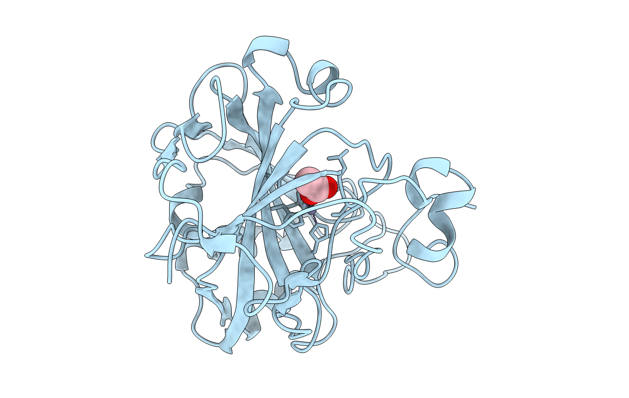
PDB ID:
1CAY
Keywords:
Title:
WILD-TYPE AND E106Q MUTANT CARBONIC ANHYDRASE COMPLEXED WITH ACETATE
Biological Source:
Source Organism(s):
Homo sapiens (Taxon ID: 9606)
Method Details:
Experimental Method:
Resolution:
2.10 Å
R-Value Observed:
0.14
Space Group:
P 1 21 1