1CAH image
Deposition Date 1992-06-25
Release Date 1993-10-31
Last Version Date 2024-02-07
Entry Detail
PDB ID:
1CAH
Keywords:
Title:
STRUCTURE OF COBALT CARBONIC ANHYDRASE COMPLEXED WITH BICARBONATE
Biological Source:
Source Organism:
Homo sapiens (Taxon ID: 9606)
Method Details:
Experimental Method:
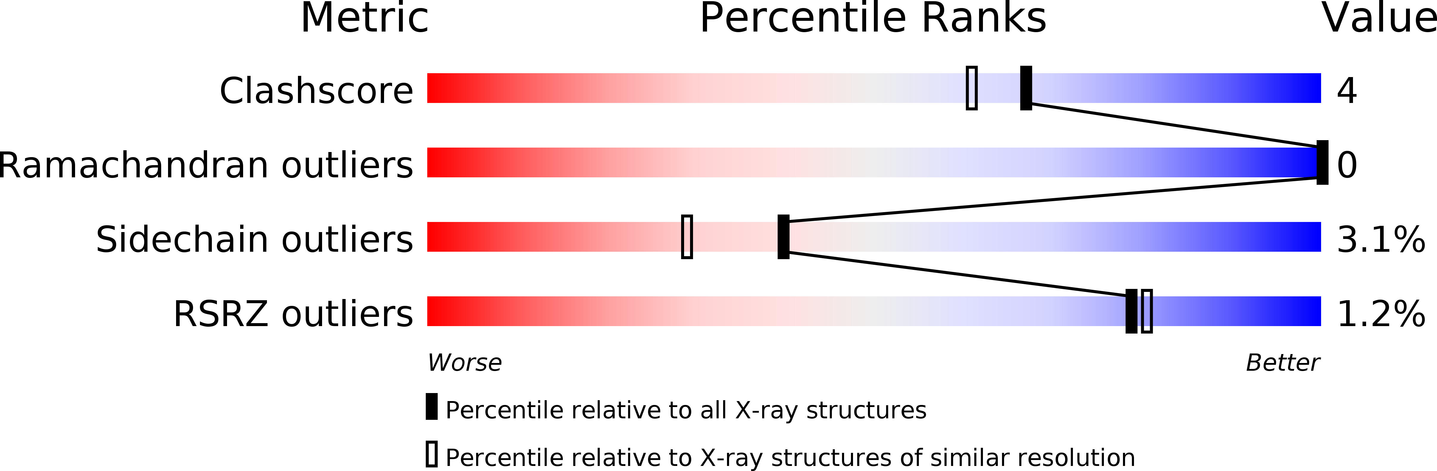
Resolution:
1.88 Å
R-Value Observed:
0.16
Space Group:
P 1 21 1
Macromolecular Entities
Structures with similar UniProt ID
Protein Blast
Polymer Type:polypeptide(L)
Molecule:CARBONIC ANHYDRASE II
Gene (Uniprot):CA2
Chain IDs:A
Chain Length:259
Number of Molecules:1
Biological Source:Homo sapiens
Primary Citation
Structure of cobalt carbonic anhydrase complexed with bicarbonate.
J.Mol.Biol. 228 1212 1218 (1992)
PMID: 1474587 DOI: 10.1016/0022-2836(92)90327-G

Abstact

The three-dimensional structure of a complex between catalytically active cobalt(II) substituted human carbonic anhydrase II and its substrate bicarbonate was determined by X-ray crystallography (1.9 A). One water molecule and two bicarbonate oxygen atoms are found at distances between 2.3 and 2.5 A from the cobalt ion in addition to the three histidyl ligands contributed by the peptide chain. The tetrahedral geometry around the metal ion in the native enzyme with a single water molecule 2.0 A from the metal is therefore lost. The geometry is difficult to classify but might best be described as distorted octahedral. The structure is suggested to represent a water-bicarbonate exchange state relevant also for native carbonic anhydrase, where the two unprotonized oxygen atoms of the substrate are bound in a carboxylate binding site and the hydroxyl group is free to move closer to the metal thereby replacing the metal-bound water molecule. A reaction mechanism based on crystallographically determined enzyme-ligand complexes is represented.

Legend

Protein

Chemical

Disease

Primary Citation of related structures