
Deposition Date
2000-03-17
Release Date
2000-09-20
Last Version Date
2024-11-06
Entry Detail
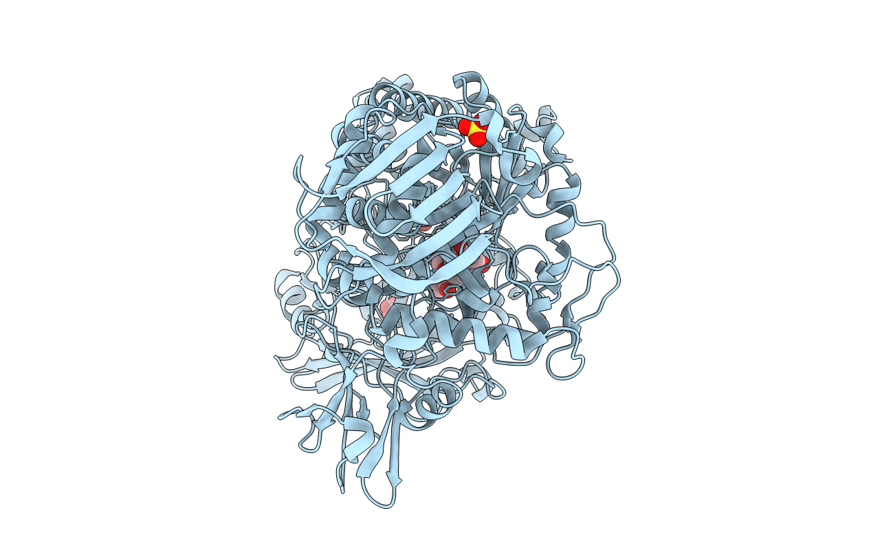
PDB ID:
1C7T
Keywords:
Title:
BETA-N-ACETYLHEXOSAMINIDASE MUTANT E540D COMPLEXED WITH DI-N ACETYL-D-GLUCOSAMINE (CHITOBIASE)
Biological Source:
Source Organism(s):
Serratia marcescens (Taxon ID: 615)
Expression System(s):
Method Details:
Experimental Method:
Resolution:
1.90 Å
R-Value Free:
0.24
R-Value Work:
0.19
Space Group:
P 21 21 2
