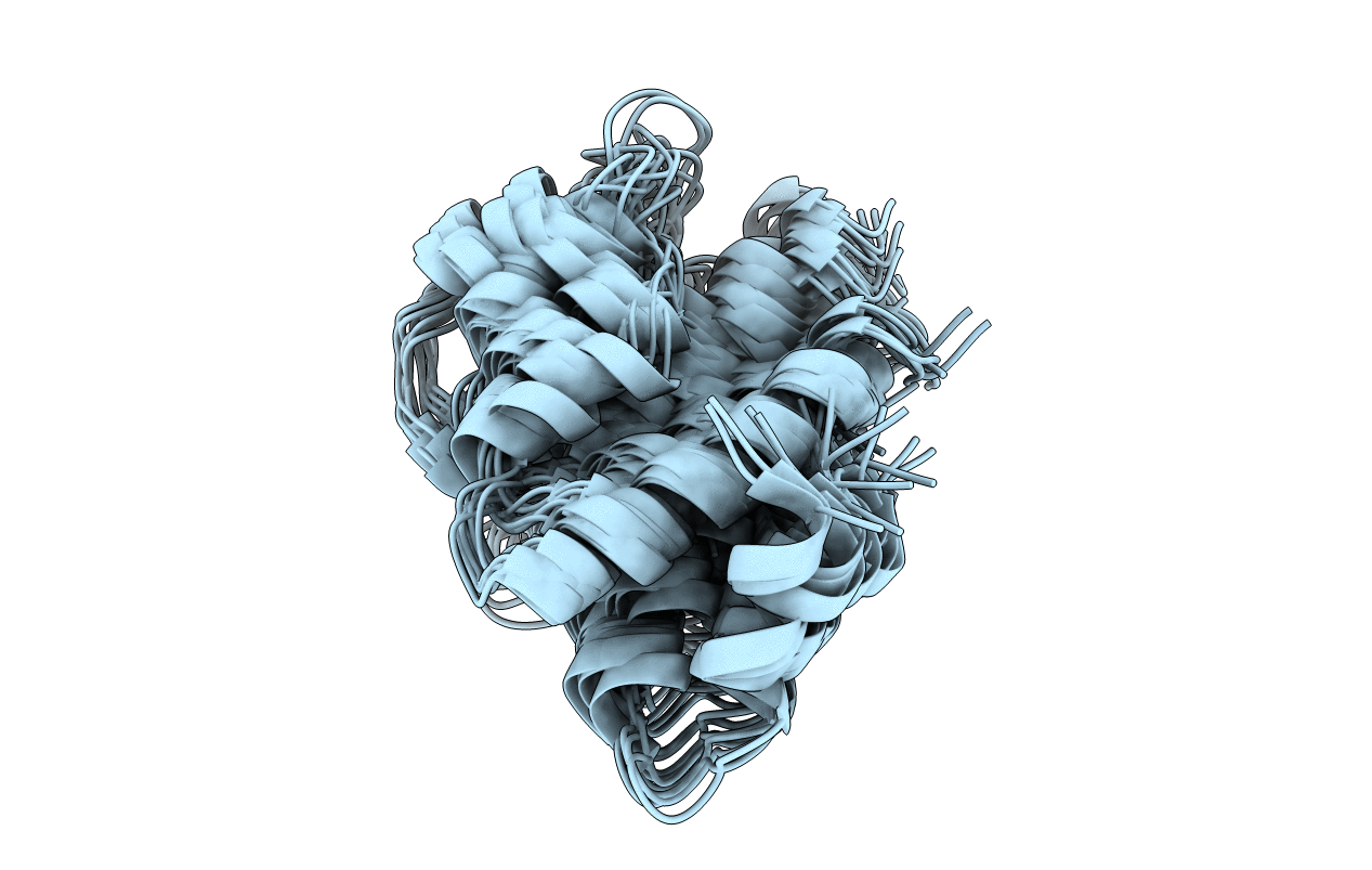
1C5A image
Deposition Date 1990-06-12
Release Date 1991-10-15
Last Version Date 2024-10-16
Entry Detail
PDB ID:
1C5A
Title:
THREE-DIMENSIONAL STRUCTURE OF PORCINE C5ADES*ARG FROM 1H NUCLEAR MAGNETIC RESONANCE DATA
Biological Source:
Source Organism:
Method Details:
Experimental Method:
Conformers Submitted:
41
Macromolecular Entities
Polymer Type:polypeptide(L)
Molecule:COMPLEMENT C5A ANAPHYLATOXIN
Gene (Uniprot):C5
Chain IDs:A
Chain Length:73
Number of Molecules:1
Biological Source:Sus scrofa domestica
Ligand Molecules
Primary Citation
Three-dimensional structure of porcine C5adesArg from 1H nuclear magnetic resonance data.
Biochemistry 29 2895 2905 (1990)
PMID: 2337573 DOI: 10.1021/bi00464a002

Abstact

Two-dimensional nuclear magnetic resonance spectra of porcine C5adesArg (73 residues) have been used to construct a list of 34 hydrogen bonds, 27 dihedral angle constraints, and 151 distance constraints, derived from nuclear Overhauser effect data. These constraints were used in restrained molecular dynamics calculations on residues 1-65 of C5a, starting from a folded structure modeled on the crystal structure of a homologous protein, C3a. Forty-one structures have been calculated, which fall into three similar families with few violations of the imposed constraints. Structures in the most populated family have a root-mean-square deviation from the average structure of 1.02 A for the C alpha atoms, with good definition of the internal residues. There is good agreement between the calculated structures and other nuclear magnetic resonance data. The structure is very similar to that recently reported for human C5a [Zuiderweg et al. (1989) Biochemistry 28, 172-185]. Some biological implications of these structures are discussed.

Legend

Protein

Chemical

Disease

Primary Citation of related structures