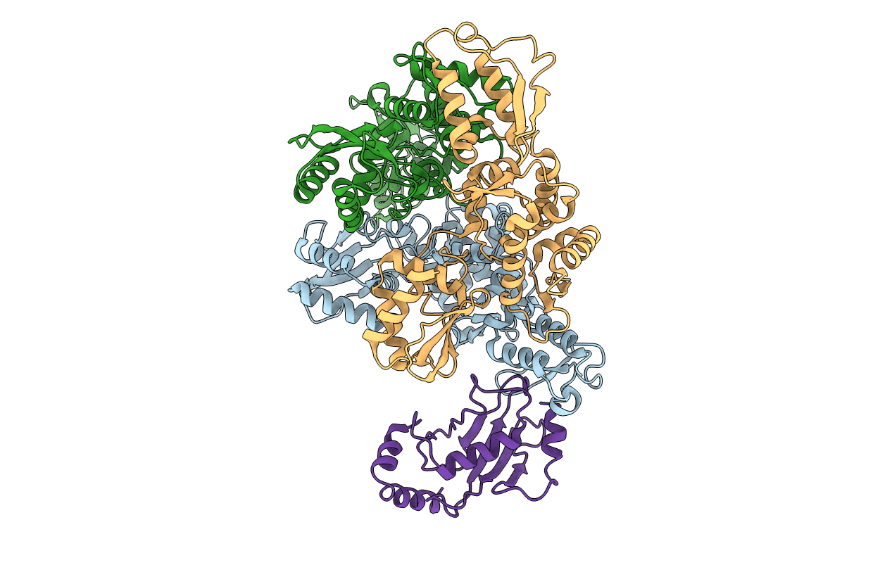
1C4Z image
Deposition Date 1999-10-14
Release Date 1999-11-17
Last Version Date 2024-02-07
Entry Detail
PDB ID:
1C4Z
Keywords:
Title:
STRUCTURE OF AN E6AP-UBCH7 COMPLEX: INSIGHTS INTO THE UBIQUITINATION PATHWAY
Biological Source:
Source Organism:
Homo sapiens (Taxon ID: 9606)
Host Organism:
Method Details:
Experimental Method:
Resolution:
2.60 Å
R-Value Free:
0.29
R-Value Work:
0.23
R-Value Observed:
0.23
Space Group:
P 21 21 21
Macromolecular Entities
Structures with similar UniProt ID
Protein Blast
Polymer Type:polypeptide(L)
Molecule:UBIQUITIN-PROTEIN LIGASE E3A
Gene (Uniprot):UBE3A
Chain IDs:A, B, C
Chain Length:358
Number of Molecules:3
Biological Source:Homo sapiens
Structures with similar UniProt ID
Protein Blast
Polymer Type:polypeptide(L)
Molecule:UBIQUITIN CONJUGATING ENZYME E2
Gene (Uniprot):UBE2L3
Chain IDs:D
Chain Length:154
Number of Molecules:1
Biological Source:Homo sapiens
Primary Citation
Structure of an E6AP-UbcH7 complex: insights into ubiquitination by the E2-E3 enzyme cascade.
Science 286 1321 1326 (1999)
PMID: 10558980 DOI: 10.1126/science.286.5443.1321

Abstact

The E6AP ubiquitin-protein ligase (E3) mediates the human papillomavirus-induced degradation of the p53 tumor suppressor in cervical cancer and is mutated in Angelman syndrome, a neurological disorder. The crystal structure of the catalytic hect domain of E6AP reveals a bilobal structure with a broad catalytic cleft at the junction of the two lobes. The cleft consists of conserved residues whose mutation interferes with ubiquitin-thioester bond formation and is the site of Angelman syndrome mutations. The crystal structure of the E6AP hect domain bound to the UbcH7 ubiquitin-conjugating enzyme (E2) reveals the determinants of E2-E3 specificity and provides insights into the transfer of ubiquitin from the E2 to the E3.

Legend

Protein

Chemical

Disease

Primary Citation of related structures