Deposition Date
1999-07-28
Release Date
2000-04-10
Last Version Date
2023-12-27
Entry Detail
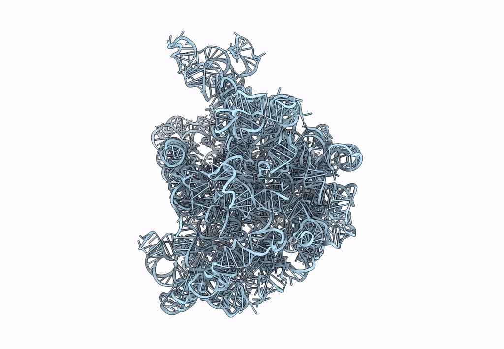
PDB ID:
1C2W
Keywords:
Title:
23S RRNA STRUCTURE FITTED TO A CRYO-ELECTRON MICROSCOPIC MAP AT 7.5 ANGSTROMS RESOLUTION
Biological Source:
Source Organism(s):
Escherichia coli (Taxon ID: 562)
Method Details:
Experimental Method:
Resolution:
7.50 Å
Aggregation State:
PARTICLE
Reconstruction Method:
SINGLE PARTICLE