1C2N image
Deposition Date 1998-04-27
Release Date 1999-03-23
Last Version Date 2024-10-30
Entry Detail
PDB ID:
1C2N
Title:
CYTOCHROME C2, NMR, 20 STRUCTURES
Biological Source:
Source Organism:
Method Details:
Experimental Method:
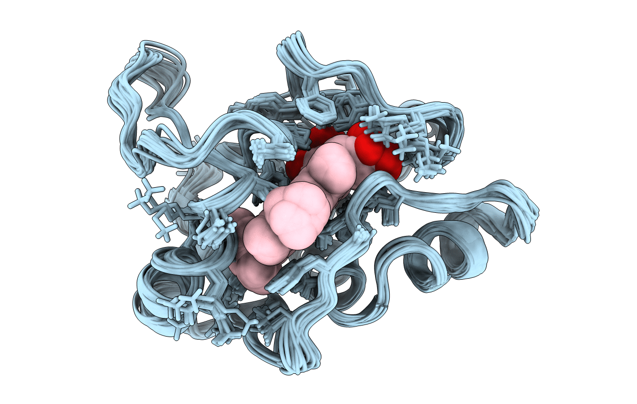
Conformers Calculated:
25
Conformers Submitted:
20
Selection Criteria:
LEAST RESTRAINT VIOLATION
Macromolecular Entities
Polymer Type:polypeptide(L)
Molecule:CYTOCHROME C2
Gene (Uniprot):cycA
Chain IDs:A
Chain Length:137
Number of Molecules:1
Biological Source:Rhodobacter capsulatus
Ligand Molecules
Primary Citation
Solution structure, rotational diffusion anisotropy and local backbone dynamics of Rhodobacter capsulatus cytochrome c2.
J.Mol.Biol. 281 341 361 (1998)
PMID: 9698552 DOI: 10.1006/jmbi.1998.1950

Abstact

The solution structure, backbone dynamics and rotational diffusion of the Rhodobacter capsulatus cytochrome c2 have been determined using heteronuclear NMR spectroscopy. In all, 1204 NOE-derived distances were used in the structure calculation to give a final ensemble with 0.59(+/-0.08) A rms deviation for the backbone atoms (C, Calpha and N) with respect to the mean coordinates. There is no major difference between the solution structure and the previously solved X-ray crystal structure (1.07(+/-0.07) A rms difference for the backbone atoms), although certain significant local structural differences have been identified. This protein contains five helical regions and a histidine-heme binding domain, connected by a series of structured loops. The orientation of the helices provides an excellent sampling of angular space and thus allows a precise characterization of the anisotropic diffusion tensor. Analysis of the hydrodynamics of the protein has been performed by interpretation of the 15N relaxation data using isotropic, axially asymmetric and fully anisotropic diffusion tensors. The protein can be shown to exhibit significant anisotropic reorientation with a diffusion tensor with principal axes values of 1.405(+/-0.031)x10(7) s-1, 1.566(+/-0.051)x10(7) s-1 and 1.829(+/-0.054)x10(7) s-1. Hydrodynamic calculations performed on the solution structure predict values of 1.399x10(7) s-1, 1.500x10(7) s-1 and 1.863x10(7) s-1 when a solvent shell of 3.5 A is included in the calculation. The optimal orientation of the diffusion tensor has been incorporated into a hybrid Lipari-Szabo type local motion-anisotropic rotational diffusion model to characterize the local mobility in the molecule. The mobility parameters thus extracted show a quantitative improvement with respect to the model-free analysis assuming isotropic reorientation; helical regions exhibit similar dynamic properties and fewer residues require more complex models of internal motion. While the molecule is essentially rigid, a tripeptide loop region (residues 101 to 103) exhibits flexibility in the range of 20 to 30 ps, which appears to be correlated with the order in the NMR solution structure.

Legend

Protein

Chemical

Disease

Primary Citation of related structures