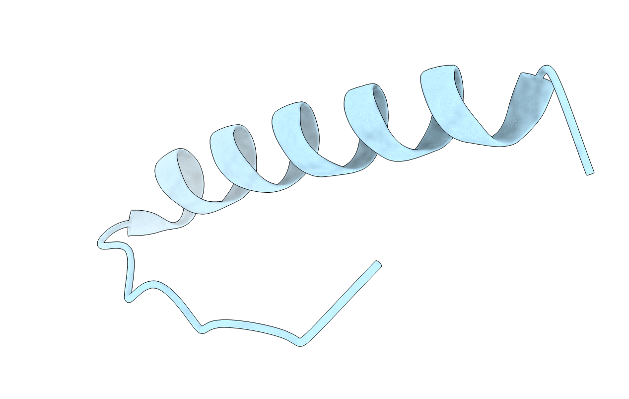
1C26 image
Deposition Date 1999-07-22
Release Date 1999-07-27
Last Version Date 2024-02-07
Entry Detail
PDB ID:
1C26
Keywords:
Title:
CRYSTAL STRUCTURE OF P53 TETRAMERIZATION DOMAIN
Biological Source:
Source Organism:
Homo sapiens (Taxon ID: 9606)
Host Organism:
Method Details:
Experimental Method:
Resolution:
1.70 Å
R-Value Work:
0.19
R-Value Observed:
0.19
Space Group:
P 4 2 2
Macromolecular Entities
Structures with similar UniProt ID
Protein Blast
Polymer Type:polypeptide(L)
Molecule:P53 TUMOR SUPPRESSOR
Gene (Uniprot):TP53
Chain IDs:A
Chain Length:32
Number of Molecules:1
Biological Source:Homo sapiens
Primary Citation
Crystal structure of the tetramerization domain of the p53 tumor suppressor at 1.7 angstroms.
Science 267 1498 1502 (1995)
PMID: 7878469

Abstact

The p53 protein is a tetrameric transcription factor that plays a central role in the prevention of neoplastic transformation. Oligomerization appears to be essential for the tumor suppressing activity of p53 because oligomerization-deficient p53 mutants cannot suppress the growth of carcinoma cell lines. The crystal structure of the tetramerization domain of p53 (residues 325 to 356) was determined at 1.7 angstrom resolution and refined to a crystallographic R factor of 19.2 percent. The monomer, which consists of a beta strand and an alpha helix, associates with a second monomer across an antiparallel beta sheet and an antiparallel helix-helix interface to form a dimer. Two of these dimers associate across a second and distinct parallel helix-helix interface to form the tetramer.

Legend

Protein

Chemical

Disease

Primary Citation of related structures