Deposition Date
1999-07-22
Release Date
1999-11-19
Last Version Date
2024-10-30
Entry Detail
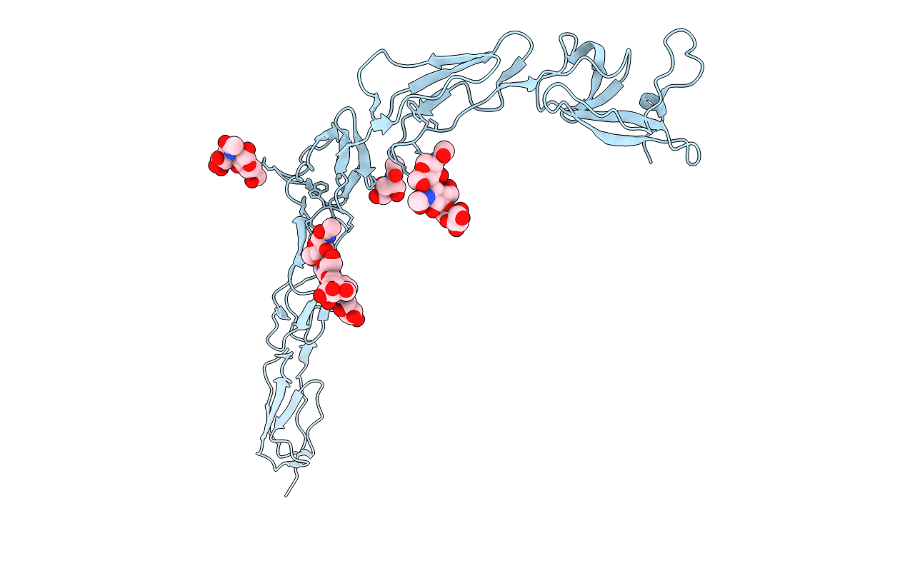
PDB ID:
1C1Z
Keywords:
Title:
CRYSTAL STRUCTURE OF HUMAN BETA-2-GLYCOPROTEIN-I (APOLIPOPROTEIN-H)
Biological Source:
Source Organism(s):
Homo sapiens (Taxon ID: 9606)
Method Details:
Experimental Method:
Resolution:
2.87 Å
R-Value Free:
0.24
R-Value Work:
0.23
R-Value Observed:
0.23
Space Group:
C 2 2 21