1C16 image
Deposition Date 1999-07-20
Release Date 2000-01-26
Last Version Date 2024-10-30
Entry Detail
PDB ID:
1C16
Keywords:
Title:
CRYSTAL STRUCTURE ANALYSIS OF THE GAMMA/DELTA T CELL LIGAND T22
Biological Source:
Source Organism:
Mus musculus (Taxon ID: 10090)
Homo sapiens (Taxon ID: 9606)
Method Details:
Experimental Method:
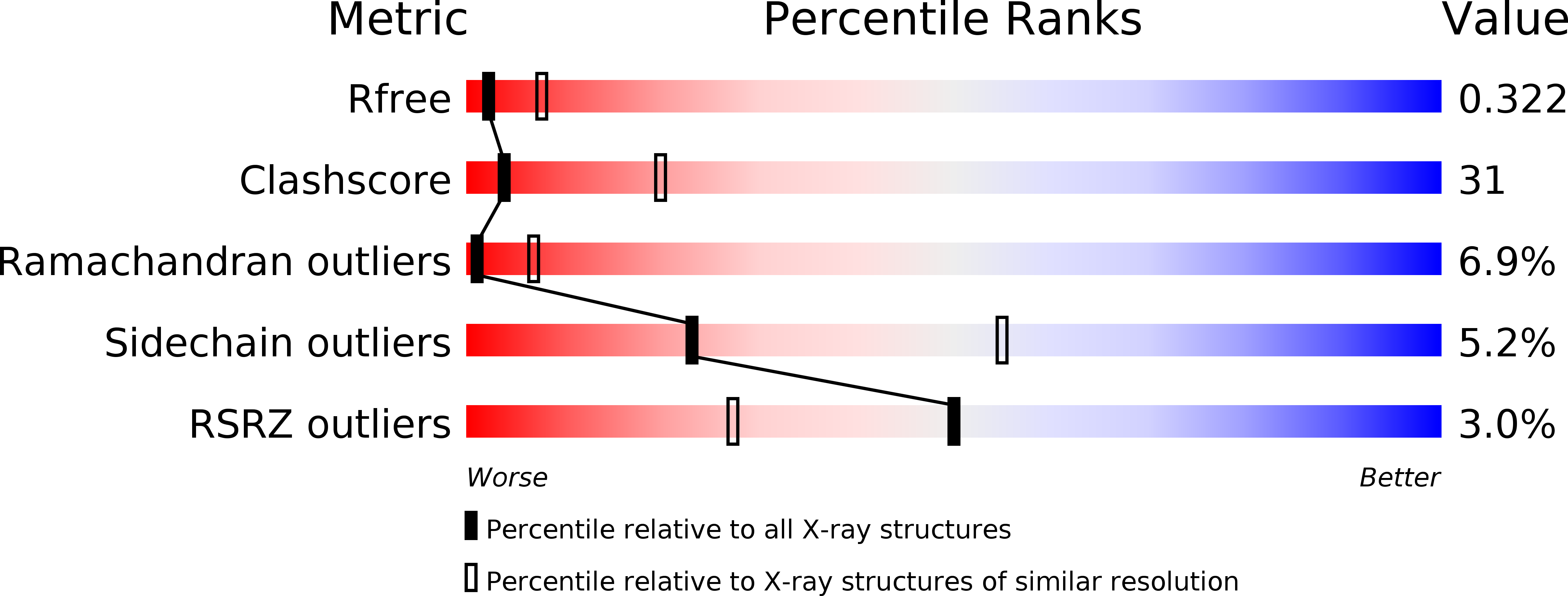
Resolution:
3.10 Å
R-Value Free:
0.33
R-Value Work:
0.28
R-Value Observed:
0.28
Space Group:
P 21 21 2
Macromolecular Entities
Structures with similar UniProt ID
Protein Blast
Polymer Type:polypeptide(L)
Molecule:MHC-LIKE PROTEIN T22
Gene (Uniprot):H2-T22
Chain IDs:A, C, E, G
Chain Length:260
Number of Molecules:4
Biological Source:Mus musculus
Structures with similar UniProt ID
Protein Blast
Polymer Type:polypeptide(L)
Molecule:PROTEIN (BETA-2-MICROGLOBULIN)
Gene (Uniprot):B2M
Chain IDs:B, D, F, H
Chain Length:99
Number of Molecules:4
Biological Source:Homo sapiens
Ligand Molecules
Primary Citation
Crystal structure of a gammadelta T cell receptor ligand T22: a truncated MHC-like fold.
Science 287 310 314 (2000)
PMID: 10634787 DOI: 10.1126/science.287.5451.310

Abstact

Murine T10 and T22 are highly related nonclassical major histocompatibility complex (MHC) class Ib proteins that bind to certain gammadelta T cell receptors (TCRs) in the absence of other components. The crystal structure of T22b at 3.1 angstroms reveals similarities to MHC class I molecules, but one side of the normal peptide-binding groove is severely truncated, which allows direct access to the beta-sheet floor. Potential gammadelta TCR-binding sites can be inferred from functional mapping of T10 and T22 point mutants and allelic variants. Thus, T22 represents an unusual variant of the MHC-like fold and indicates that gammadelta and alphabeta TCRs interact differently with their respective MHC ligands.

Legend

Protein

Chemical

Disease

Primary Citation of related structures
Feedback Form
Name
Email
Institute
Feedback