1C0A image
Entry Detail
PDB ID:
1C0A
Keywords:
Title:
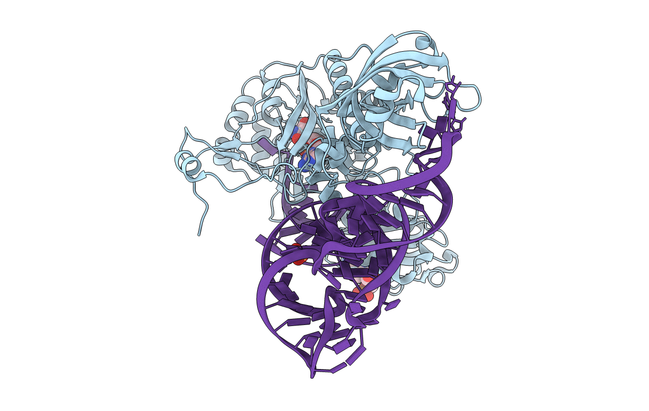
CRYSTAL STRUCTURE OF THE E. COLI ASPARTYL-TRNA SYNTHETASE : TRNAASP : ASPARTYL-ADENYLATE COMPLEX
Biological Source:
Source Organism:
Host Organism:
PDB Version:
Deposition Date:
1999-07-15
Release Date:
1999-11-23
Method Details:
Experimental Method:
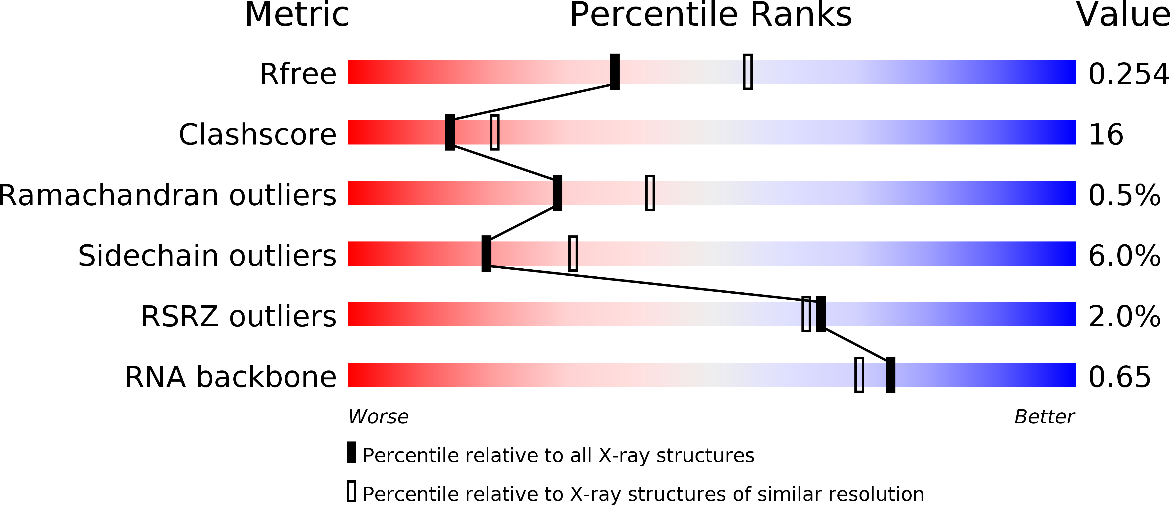
Resolution:
2.40 Å
R-Value Free:
0.24
R-Value Work:
0.20
R-Value Observed:
0.20
Space Group:
P 43 21 2
Macromolecular Entities
Polymer Type:polypeptide(L)
Description:ASPARTYL TRNA SYNTHETASE
Chain IDs:B (auth: A)
Chain Length:585
Number of Molecules:1
Biological Source:Escherichia coli
Polymer Type:polyribonucleotide
Description:ASPARTYL TRNA
Chain IDs:A (auth: B)
Chain Length:77
Number of Molecules:1
Biological Source:Escherichia coli
Modified Residue
Compound ID Chain ID Parent Comp ID Details 2D Image
4SU A U 4-THIOURIDINE-5'-MONOPHOSPHATE
5MU A U 5-METHYLURIDINE 5'-MONOPHOSPHATE
G7M A G N7-METHYL-GUANOSINE-5'-MONOPHOSPHATE
H2U A U 5,6-DIHYDROURIDINE-5'-MONOPHOSPHATE
PSU A U PSEUDOURIDINE-5'-MONOPHOSPHATE
QUO A G ?
Primary Citation
Synthesis of aspartyl-tRNA(Asp) in Escherichia coli--a snapshot of the second step.
EMBO J. 18 6532 6541 (1999)
PMID: 10562565 DOI: 10.1093/emboj/18.22.6532

Abstact

The 2.4 A crystal structure of the Escherichia coli aspartyl-tRNA synthetase (AspRS)-tRNA(Asp)-aspartyl-adenylate complex shows the two substrates poised for the transfer of the aspartic acid moiety from the adenylate to the 3'-hydroxyl of the terminal adenosine of the tRNA. A general molecular mechanism is proposed for the second step of the aspartylation reaction that accounts for the observed conformational changes, notably in the active site pocket. The stabilization of the transition state is mediated essentially by two amino acids: the class II invariant arginine of motif 2 and the eubacterial-specific Gln231, which in eukaryotes and archaea is replaced by a structurally non-homologous serine. Two archetypal RNA-protein modes of interactions are observed: the anticodon stem-loop, including the wobble base Q, binds to the N-terminal beta-barrel domain through direct protein-RNA interactions, while the binding of the acceptor stem involves both direct and water-mediated hydrogen bonds in an original recognition scheme.

Legend

Protein

Chemical

Disease

Primary Citation of related structures