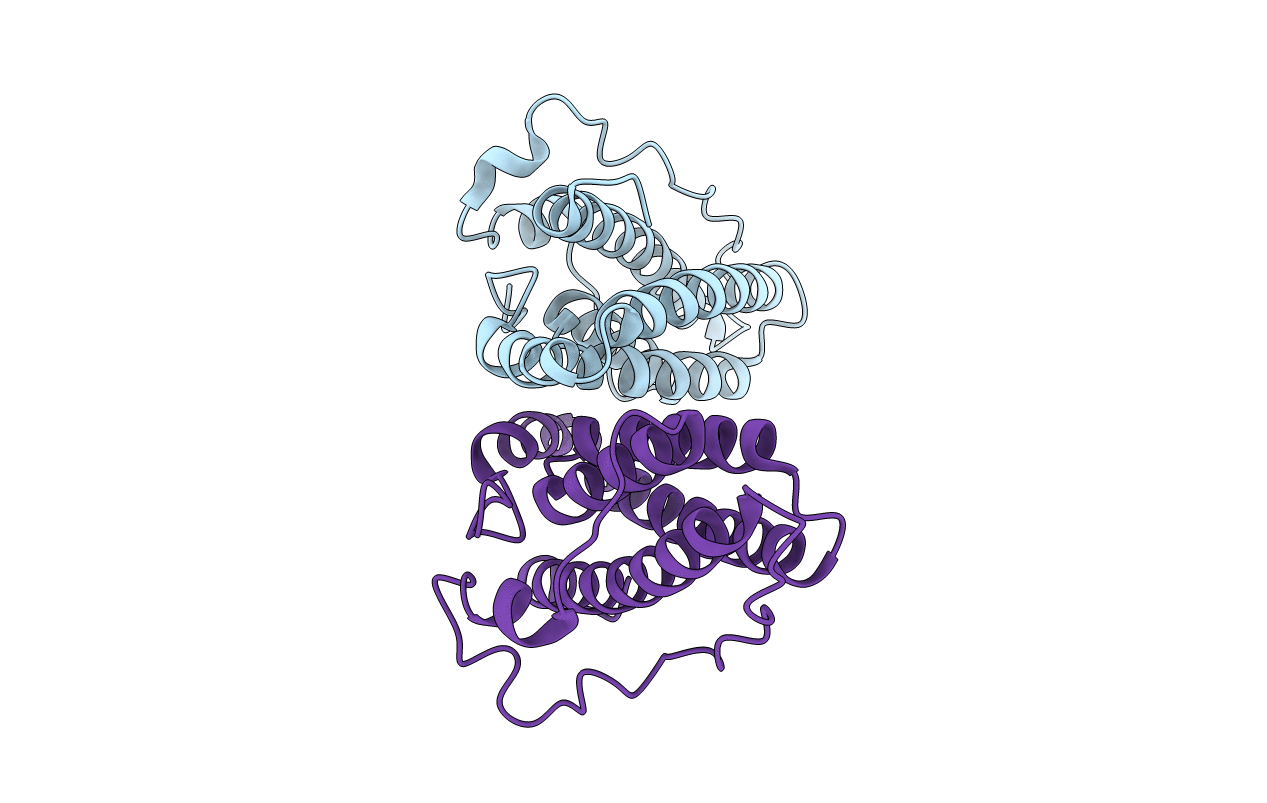
1C02 image
Deposition Date 1999-07-14
Release Date 2000-01-15
Last Version Date 2024-02-07
Entry Detail
PDB ID:
1C02
Keywords:
Title:
CRYSTAL STRUCTURE OF YEAST YPD1P
Biological Source:
Source Organism:
Host Organism:
Method Details:
Experimental Method:
Resolution:
1.80 Å
R-Value Free:
0.24
R-Value Work:
0.20
R-Value Observed:
0.20
Space Group:
P 43 21 2
Macromolecular Entities
Polymer Type:polypeptide(L)
Molecule:PHOSPHOTRANSFERASE YPD1P
Gene (Uniprot):YPD1
Chain IDs:A, B
Chain Length:166
Number of Molecules:2
Biological Source:Saccharomyces cerevisiae
Primary Citation
Insights into eukaryotic multistep phosphorelay signal transduction revealed by the crystal structure of Ypd1p from Saccharomyces cerevisiae.
J.Mol.Biol. 293 753 761 (1999)
PMID: 10543964 DOI: 10.1006/jmbi.1999.3215

Abstact

"Two-component" phosphorelay signal transduction systems constitute a potential target for antibacterial and antifungal agents, since they are found exclusively in prokaryotes and lower eukaryotes (yeast, fungi, slime mold, and plants) but not in mammalian organisms. Saccharomyces cerevisiae Ypd1p, a key intermediate in the osmosensing multistep phosphorelay signal transduction, catalyzes the phosphoryl group transfer between response regulators. Its 1.8 A structure, representing the first example of a eukaryotic phosphorelay protein, contains a four-helix bundle as in the HPt domain of Escherichia coli ArcB sensor kinase. However, Ypd1p has a 44-residue insertion between the last two helices of the helix bundle. The side-chain of His64, the site of phosphorylation, protrudes into the solvent. The structural resemblance between Ypd1p and ArcB HPt domain suggests that both prokaryotes and lower eukaryotes utilize the same basic protein fold for phosphorelay signal transduction. This study sheds light on the best characterized eukaryotic phosphorelay system.

Legend

Protein

Chemical

Disease

Primary Citation of related structures