Deposition Date
1998-11-03
Release Date
1998-11-11
Last Version Date
2024-11-06
Entry Detail
PDB ID:
1BZQ
Keywords:
Title:
COMPLEX OF A DROMEDARY SINGLE-DOMAIN VHH ANTIBODY FRAGMENT WITH RNASE A
Biological Source:
Source Organism(s):
Camelus dromedarius (Taxon ID: 9838)
Bos taurus (Taxon ID: 9913)
Bos taurus (Taxon ID: 9913)
Expression System(s):
Method Details:
Experimental Method:
Resolution:
2.80 Å
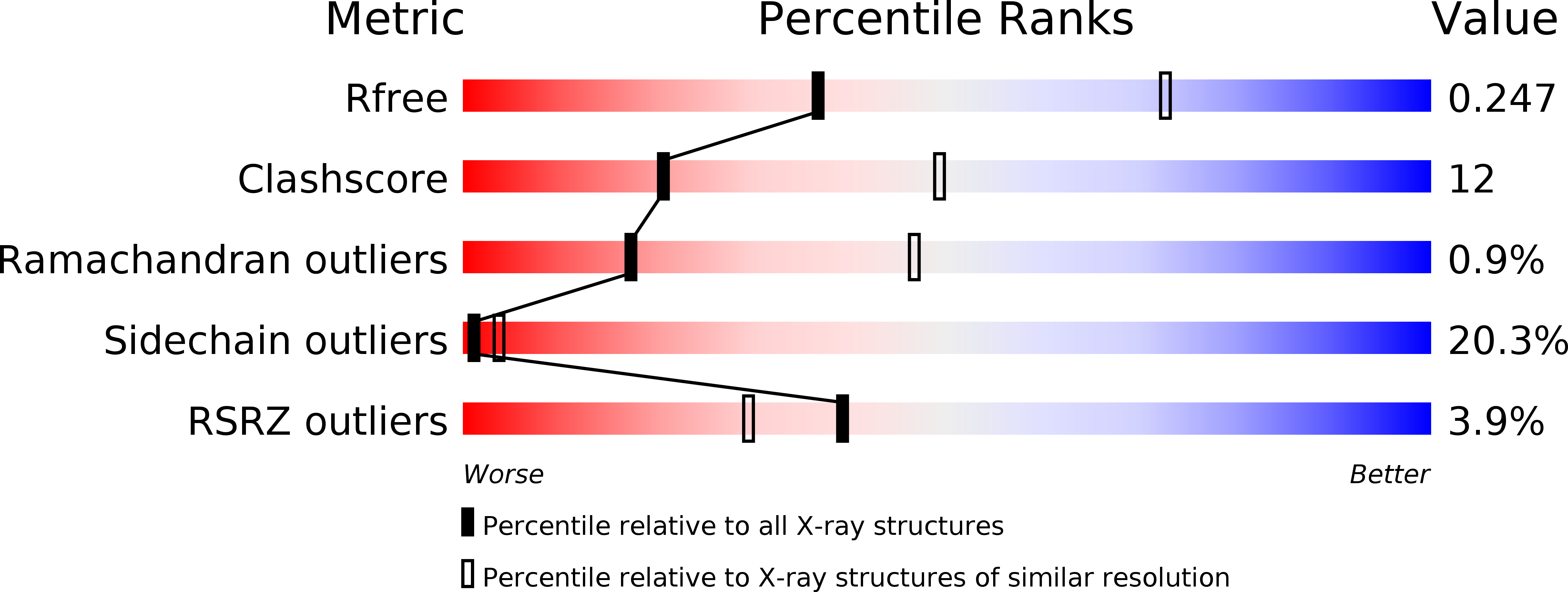
R-Value Free:
0.28
R-Value Work:
0.22
Space Group:
P 1