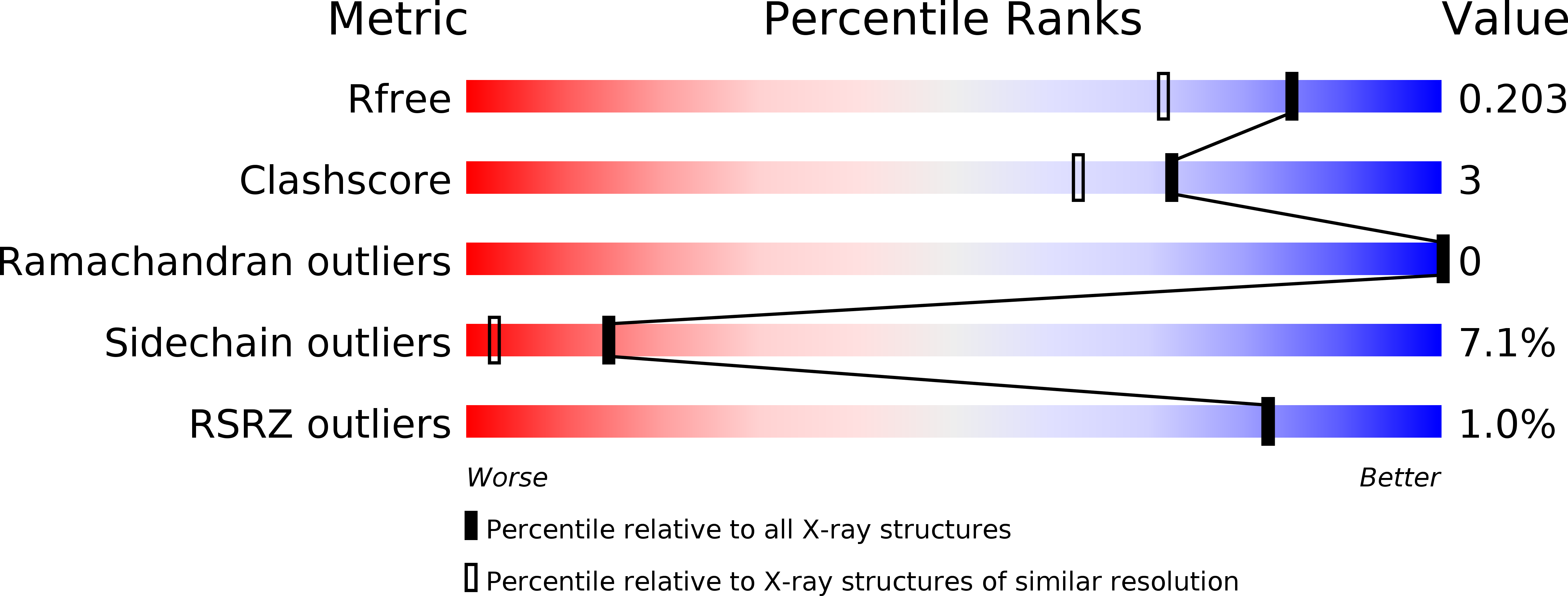
1BZ1 image
Deposition Date 1998-11-05
Release Date 1998-11-11
Last Version Date 2024-04-03
Entry Detail
PDB ID:
1BZ1
Title:
HEMOGLOBIN (ALPHA + MET) VARIANT
Biological Source:
Source Organism:
Homo sapiens (Taxon ID: 9606)
Host Organism:
Method Details:
Experimental Method:
Resolution:
1.59 Å
R-Value Free:
0.22
R-Value Work:
0.17
Space Group:
P 1 21 1
Macromolecular Entities
Structures with similar UniProt ID
Protein Blast
Polymer Type:polypeptide(L)
Molecule:PROTEIN (HEMOGLOBIN ALPHA CHAIN)
Gene (Uniprot):HBA1, HBA2
Chain IDs:A, C
Chain Length:142
Number of Molecules:2
Biological Source:Homo sapiens
Structures with similar UniProt ID
Protein Blast
Polymer Type:polypeptide(L)
Molecule:PROTEIN (HEMOGLOBIN BETA CHAIN)
Gene (Uniprot):HBB
Chain IDs:B, D
Chain Length:146
Number of Molecules:2
Biological Source:Homo sapiens
Ligand Molecules
Primary Citation
Structural and functional properties of human hemoglobins reassembled after synthesis in Escherichia coli.
Biochemistry 38 1040 1049 (1999)
PMID: 9894000 DOI: 10.1021/bi981986g

Abstact

Human hemoglobin produced in the Escherichia coli coexpression system of Hernan et al. [(1992) Biochemistry 31, 8619-8628] has been transformed into a functionally homogeneous protein whose properties closely approximate those of normal hemoglobin A. Both of the alpha and beta chains of this hemoglobin contain a valine-methionine substitution at position 1 in order to accommodate the difference in specificity of the protein-processing enzymes of procaryotes. Despite extensive purification, functional homogeneity of the E. coli expressed hemoglobin was achieved only by the complete disassembly of the hemoglobin into its component alpha and beta globins and their reassembly in the presence of hemin. The kinetics of CO combination and the thermodynamics of O2 binding and cooperativity of the reassembled alphaV1M-betaV1M hemoglobin closely approximate those of HbA. The alpha globin obtained from the E. coli expressed hemoglobin was also combined with normal human beta chains and hemin to form the alphaV1M variant. The alpha+M variant of HbA, in which the normal N-terminal valine of the alpha chains is preceded by a methionine residue, was prepared by the same procedure. The kinetics of the reactions of CO with the alphaV1M and alpha+M variants are similar to those for HbA. The equilibria of oxygen binding to alphaV1M and HbA are similar whereas alpha+M exhibits a significantly higher oxygen affinity. The three-dimensional structures of alphaV1M and alpha+M offer an explanation for the latter affinity difference. Although the structures of alphaV1M and HbA, which have been determined by X-ray crystallography, are virtually indistinguishable except at the N-terminal residues, that of alpha+M indicates the displacement of a solvent molecule, possibly a chloride ion, from arginine 141alpha. Such an alteration in an anion binding site could result in increased oxygen affinity.

Legend

Protein

Chemical

Disease

Primary Citation of related structures