Deposition Date
1998-10-19
Release Date
1999-10-19
Last Version Date
2023-12-27
Entry Detail
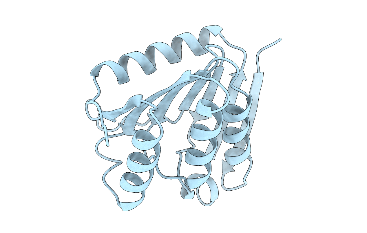
PDB ID:
1BYR
Keywords:
Title:
CRYSTAL STRUCTURE OF A PHOSPHOLIPASE D FAMILY MEMBER, NUC FROM SALMONELLA TYPHIMURIUM
Biological Source:
Source Organism(s):
Salmonella typhimurium (Taxon ID: 602)
Expression System(s):
Method Details:
Experimental Method:
Resolution:
2.00 Å
R-Value Free:
0.25
R-Value Work:
0.19
R-Value Observed:
0.19
Space Group:
P 43 21 2