Deposition Date
1998-10-19
Release Date
1999-10-19
Last Version Date
2023-08-09
Entry Detail
Biological Source:
Source Organism(s):
Silene latifolia subsp. alba (Taxon ID: 52853)
Expression System(s):
Method Details:
Experimental Method:
Resolution:
1.75 Å
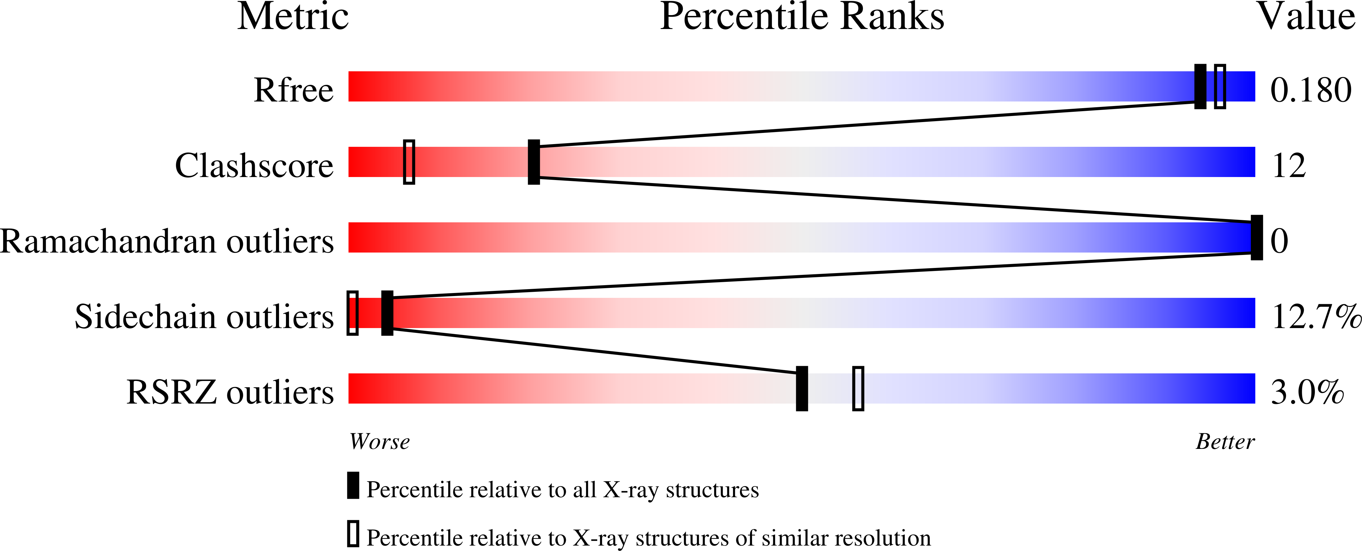
R-Value Free:
0.19
R-Value Work:
0.18
R-Value Observed:
0.18
Space Group:
P 43 21 2