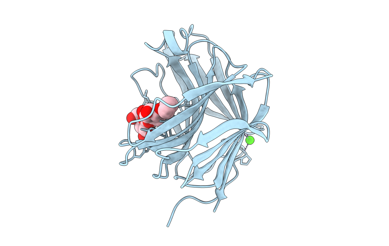
1BYH image
Deposition Date 1992-12-31
Release Date 1993-10-31
Last Version Date 2024-10-30
Entry Detail
PDB ID:
1BYH
Keywords:
Title:
MOLECULAR AND ACTIVE-SITE STRUCTURE OF A BACILLUS (1-3,1-4)-BETA-GLUCANASE
Biological Source:
Source Organism:
Method Details:
Experimental Method:
Resolution:
2.80 Å
R-Value Work:
0.16
R-Value Observed:
0.16
Space Group:
P 21 21 21
Macromolecular Entities
Structures with similar UniProt ID
Protein Blast
Polymer Type:polypeptide(L)
Molecule:HYBRID
Chain IDs:A
Chain Length:214
Number of Molecules:1
Biological Source:synthetic construct
Peptide-like Molecules
PRD_900005
Primary Citation
Molecular and active-site structure of a Bacillus 1,3-1,4-beta-glucanase.
Proc.Natl.Acad.Sci.USA 90 5287 5291 (1993)
PMID: 8099449 DOI: 10.1073/pnas.90.11.5287

Abstact

The three-dimensional structure of the hybrid Bacillus 1,3-1,4-beta-glucanase (beta-glucanase; 1,3-1,4-beta-D-glucan 4-glucanohydrolase, lichenase, EC 3.2.1.73) designated H(A16-M) was determined by x-ray crystallography at a resolution of 2.0 A and refined to an R value of 16.4% using stereochemical restraints. The protein molecule consists mainly of two seven-stranded antiparallel beta-pleated sheets arranged atop each other to form a compact, sandwich-like structure. A channel crossing one side of the protein molecule accommodates an inhibitor, 3,4-epoxybutyl beta-D-cellobioside, which binds covalently to the side chain of Glu-105, as seen in a crystal structure analysis at 2.8-A resolution of the protein-inhibitor complex (R = 16.8%). That Glu-105 may be indispensible for enzyme catalysis by H(A16-M) is suggested by site-directed mutagenesis of this residue, which inevitably leads to an inactive enzyme.

Legend

Protein

Chemical

Disease

Primary Citation of related structures