Deposition Date
1998-10-27
Release Date
1999-10-24
Last Version Date
2024-11-13
Entry Detail
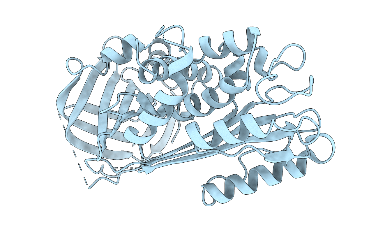
PDB ID:
1BY7
Keywords:
Title:
HUMAN PLASMINOGEN ACTIVATOR INHIBITOR-2. LOOP (66-98) DELETION MUTANT
Biological Source:
Source Organism(s):
Homo sapiens (Taxon ID: 9606)
Expression System(s):
Method Details:
Experimental Method:
Resolution:
2.00 Å
R-Value Free:
0.26
R-Value Work:
0.20
Space Group:
C 1 2 1