1BY5 image
Deposition Date 1998-10-23
Release Date 1999-01-13
Last Version Date 2023-11-15
Entry Detail
PDB ID:
1BY5
Title:
FHUA FROM E. COLI, WITH ITS LIGAND FERRICHROME
Biological Source:
Source Organism:
Method Details:
Experimental Method:
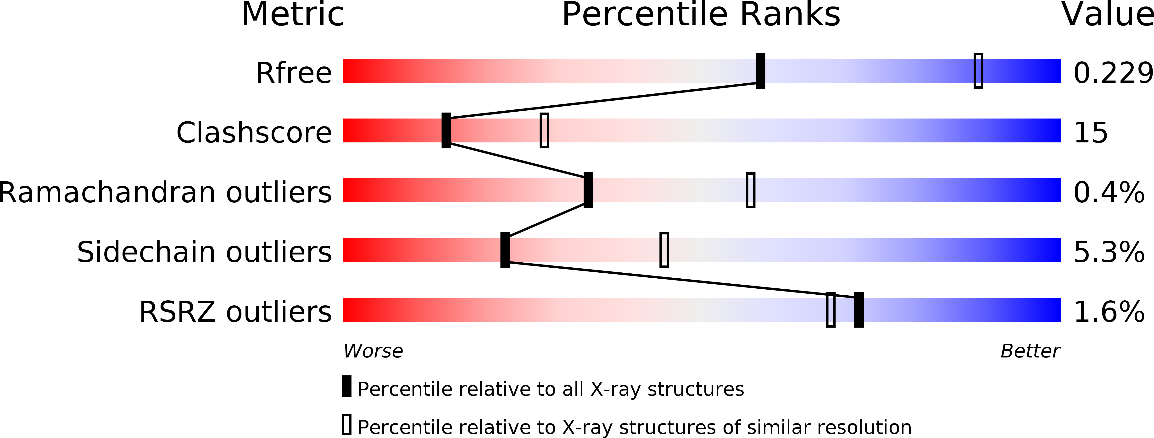
Resolution:
2.60 Å
R-Value Free:
0.22
R-Value Work:
0.18
R-Value Observed:
0.18
Space Group:
C 1 2 1
Macromolecular Entities
Structures with similar UniProt ID
Protein Blast
Polymer Type:polypeptide(L)
Molecule:FERRIC HYDROXAMATE UPTAKE PROTEIN
Chain IDs:A
Chain Length:714
Number of Molecules:1
Biological Source:Escherichia coli
Polymer Type:polypeptide(L)
Molecule:FERRICHROME
Chain IDs:B
Chain Length:6
Number of Molecules:1
Biological Source:Ustilago sphaerogena
Modified Residue
Compound ID Chain ID Parent Comp ID Details 2D Image
AHO B ALA N-ACETYL-N-HYDROXY-L-ORNITHINE
Primary Citation
Transmembrane signaling across the ligand-gated FhuA receptor: crystal structures of free and ferrichrome-bound states reveal allosteric changes.
Cell(Cambridge,Mass.) 95 771 778 (1998)
PMID: 9865695 DOI: 10.1016/S0092-8674(00)81700-6

Abstact

FhuA protein facilitates ligand-gated transport of ferrichrome-bound iron across Escherichia coli outer membranes. X-ray analysis at 2.7 A resolution reveals two distinct conformations in the presence and absence of ferrichrome. The monomeric protein consists of a hollow, 22-stranded, antiparallel beta barrel (residues 160-714), which is obstructed by a plug (residues 19-159). The binding site of ferrichrome, an aromatic pocket near the cell surface, undergoes minor changes upon association with the ligand. These are propagated and amplified across the plug, eventually resulting in substantially different protein conformations at the periplasmic face. Our findings reveal the mechanism of signal transmission and suggest how the energy-transducing TonB complex senses ligand binding.

Legend

Protein

Chemical

Disease

Primary Citation of related structures