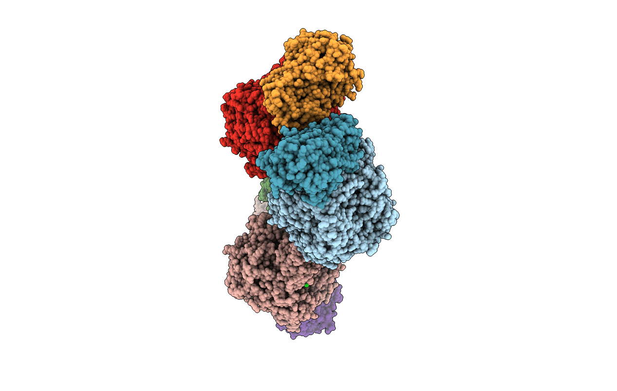
1BXR image
Deposition Date 1998-10-08
Release Date 1999-04-20
Last Version Date 2024-05-22
Entry Detail
PDB ID:
1BXR
Title:
STRUCTURE OF CARBAMOYL PHOSPHATE SYNTHETASE COMPLEXED WITH THE ATP ANALOG AMPPNP
Biological Source:
Source Organism:
Escherichia coli (Taxon ID: 562)
Host Organism:
Method Details:
Experimental Method:
Resolution:
2.10 Å
R-Value Work:
0.19
Space Group:
P 21 21 21
Macromolecular Entities
Structures with similar UniProt ID
Protein Blast
Polymer Type:polypeptide(L)
Molecule:CARBAMOYL-PHOSPHATE SYNTHASE
Gene (Uniprot):carB
Chain IDs:A, C, E, G
Chain Length:1073
Number of Molecules:4
Biological Source:Escherichia coli
Structures with similar UniProt ID
Protein Blast
Polymer Type:polypeptide(L)
Molecule:CARBAMOYL-PHOSPHATE SYNTHASE
Gene (Uniprot):carA
Chain IDs:B, D, F, H
Chain Length:382
Number of Molecules:4
Biological Source:Escherichia coli
Primary Citation
Carbamoyl phosphate synthetase: closure of the B-domain as a result of nucleotide binding.
Biochemistry 38 2347 2357 (1999)
PMID: 10029528 DOI: 10.1021/bi982517h

Abstact

Carbamoyl phosphate synthetase (CPS) catalyzes the production of carbamoyl phosphate which is subsequently employed in the metabolic pathways responsible for the synthesis of pyrimidine nucleotides or arginine. The catalytic mechanism of the enzyme occurs through three highly reactive intermediates: carboxyphosphate, ammonia, and carbamate. As isolated from Escherichia coli, CPS is an alpha, beta-heterodimeric protein with its three active sites separated by nearly 100 A. In addition, there are separate binding sites for the allosteric regulators, ornithine, and UMP. Given the sizable distances between the three active sites and the allosteric-binding pockets, it has been postulated that domain movements play key roles for intramolecular communication. Here we describe the structure of CPS from E. coli where, indeed, such a domain movement has occurred in response to nucleotide binding. Specifically, the protein was crystallized in the presence of a nonhydrolyzable analogue, AMPPNP, and its structure determined to 2.1 A resolution by X-ray crystallographic analysis. The B-domain of the carbamoyl phosphate synthetic component of the large subunit closes down over the active-site pocket such that some atoms move by more than 7 A relative to that observed in the original structure. The trigger for this movement resides in the hydrogen-bonding interactions between two backbone amide groups (Gly 721 and Gly 722) and the beta- and gamma-phosphate groups of the nucleotide triphosphate. Gly 721 and Gly 722 are located in a Type III' reverse turn, and this type of secondary structural motif is also observed in D-alanine:D-alanine ligase and glutathione synthetase, both of which belong to the "ATP-grasp" superfamily of proteins. Details concerning the geometries of the two active sites contained within the large subunit of CPS are described.

Legend

Protein

Chemical

Disease

Primary Citation of related structures