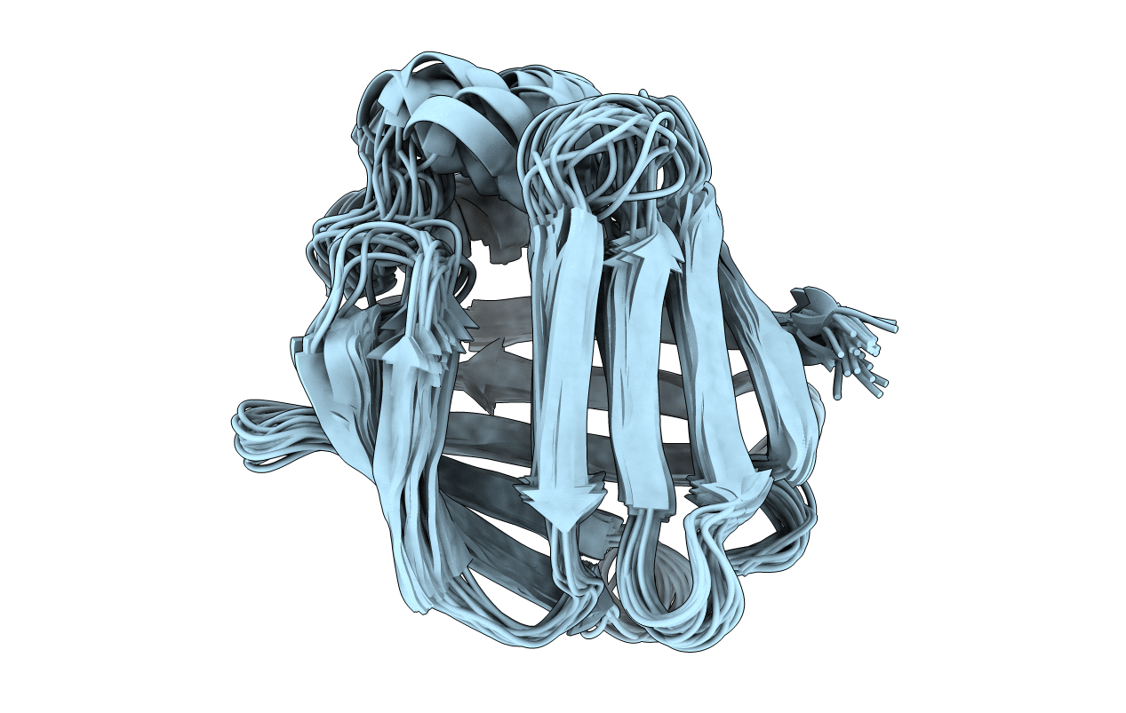
1BWY image
Deposition Date 1998-09-29
Release Date 1998-10-07
Last Version Date 2024-04-10
Entry Detail
PDB ID:
1BWY
Title:
NMR STUDY OF BOVINE HEART FATTY ACID BINDING PROTEIN
Biological Source:
Source Organism:
Bos taurus (Taxon ID: 9913)
Host Organism:
Method Details:
Experimental Method:
Conformers Calculated:
100
Conformers Submitted:
25
Selection Criteria:
LOWEST VIOLATION OF EXPERIMENTAL DISTANCE CONSTRAINTS
Macromolecular Entities
Polymer Type:polypeptide(L)
Molecule:PROTEIN (HEART FATTY ACID BINDING PROTEIN)
Gene (Uniprot):FABP3
Chain IDs:A
Chain Length:132
Number of Molecules:1
Biological Source:Bos taurus
Ligand Molecules
Primary Citation
Three-dimensional structure of bovine heart fatty-acid-binding protein with bound palmitic acid, determined by multidimensional NMR spectroscopy.
Eur.J.Biochem. 230 266 280 (1995)
PMID: 7601110 DOI: 10.1111/j.1432-1033.1995.tb20560.x

Abstact

The three-dimensional structure of the holo form of recombinant cellular bovine heart fatty-acid-binding protein (H-FABPc), a polypeptide of 133 amino acid residues with a molecular mass of 15 kDa, has been determined by multidimensional homonuclear and heteronuclear NMR spectroscopy applied to uniformly 15N-labeled and unlabeled protein. A nearly complete set of 1H and 15N chemical shift assignments was obtained. A total of 2329 intramolecular distance constraints and 42 side-chain chi 1 dihedral-angle constraints were derived from cross-relaxation and J coupling information. 3D nuclear Overhauser enhancement and exchange spectroscopy combined with heteronuclear multiple-quantum coherence (NOESY-HMQC) experiments, performed on a sample of uniformly 13C-labeled palmitic acid bound to unlabeled cellular heart fatty-acid-binding protein revealed 10 intermolecular contacts that determine the orientation of the bound fatty acid. An ensemble of protein conformations was calculated with the distance-geometry algorithm for NMR applications (DIANA) using the redundant dihedral-angle constraint (REDAC) strategy. After docking the fatty acid into the protein, the protein-ligand arrangement was subject to distance-restrained energy minimization. The overall conformation of the protein is a beta-barrel consisting of 10 antiparallel beta-strands which form two nearly orthogonal beta-sheets of five strands each. Two short helices form a helix-turn-helix motif in the N-terminal region of the polypeptide chain. The palmitic acid is bound within the protein in a U-shaped conformation close to the two helices. The obtained solution structure of the protein is consistent with a number of fatty-acid-binding-protein crystal structures.

Legend

Protein

Chemical

Disease

Primary Citation of related structures