Deposition Date
1998-09-28
Release Date
1999-01-20
Last Version Date
2024-10-09
Entry Detail
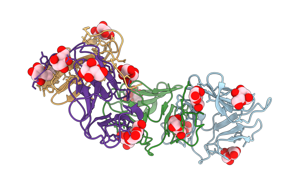
PDB ID:
1BWU
Keywords:
Title:
MANNOSE-SPECIFIC AGGLUTININ (LECTIN) FROM GARLIC (ALLIUM SATIVUM) BULBS COMPLEXED WITH ALPHA-D-MANNOSE
Biological Source:
Source Organism(s):
Allium sativum (Taxon ID: 4682)
Method Details:
Experimental Method:
Resolution:
2.80 Å
R-Value Free:
0.27
R-Value Work:
0.22
R-Value Observed:
0.22
Space Group:
C 1 2 1