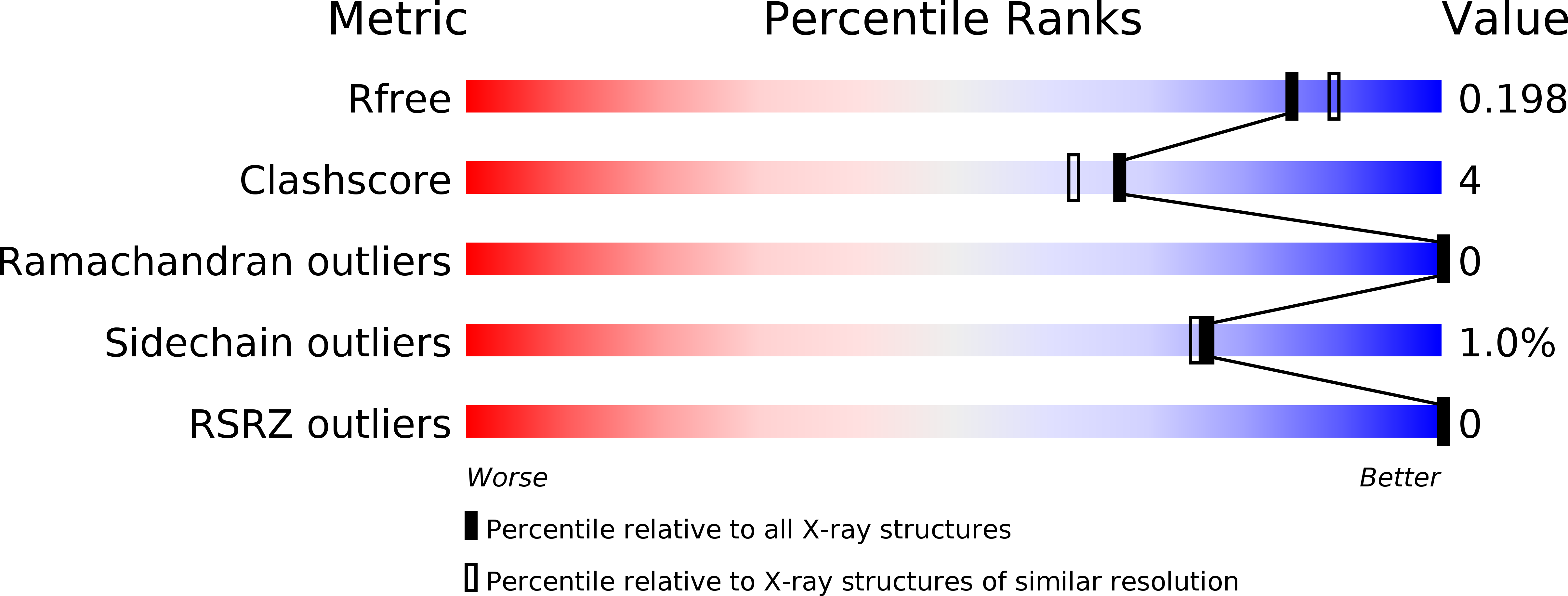
1BVW image
Deposition Date 1998-09-19
Release Date 1999-09-18
Last Version Date 2024-10-30
Entry Detail
PDB ID:
1BVW
Keywords:
Title:
CELLOBIOHYDROLASE II (CEL6A) FROM HUMICOLA INSOLENS
Biological Source:
Source Organism:
Host Organism:
Method Details:
Experimental Method:
Resolution:
1.92 Å
R-Value Free:
0.21
R-Value Work:
0.14
Space Group:
P 1 21 1
Macromolecular Entities
Polymer Type:polypeptide(L)
Molecule:PROTEIN (CELLOBIOHYDROLASE II)
Gene (Uniprot):cel6A
Chain IDs:A
Chain Length:360
Number of Molecules:1
Biological Source:Humicola insolens
Modified Residue
Compound ID Chain ID Parent Comp ID Details 2D Image
ASN A ASN GLYCOSYLATION SITE
SER A SER GLYCOSYLATION SITE
THR A THR GLYCOSYLATION SITE
Primary Citation
Crystal structure of the catalytic core domain of the family 6 cellobiohydrolase II, Cel6A, from Humicola insolens, at 1.92 A resolution.
Biochem.J. 337 297 304 (1999)
PMID: 9882628 DOI: 10.1042/0264-6021:3370297

Abstact

The three-dimensional structure of the catalytic core of the family 6 cellobiohydrolase II, Cel6A (CBH II), from Humicola insolens has been determined by X-ray crystallography at a resolution of 1.92 A. The structure was solved by molecular replacement using the homologous Trichoderma reesei CBH II as a search model. The H. insolens enzyme displays a high degree of structural similarity with its T. reesei equivalent. The structure features both O- (alpha-linked mannose) and N-linked glycosylation and a hexa-co-ordinate Mg2+ ion. The active-site residues are located within the enclosed tunnel that is typical for cellobiohydrolase enzymes and which may permit a processive hydrolysis of the cellulose substrate. The close structural similarity between the two enzymes implies that kinetics and chain-end specificity experiments performed on the H. insolens enzyme are likely to be applicable to the homologous T. reesei enzyme. These cast doubt on the description of cellobiohydrolases as exo-enzymes since they demonstrated that Cel6A (CBH II) shows no requirement for non-reducing chain-ends, as had been presumed. There is no crystallographic evidence in the present structure to support a mechanism involving loop opening, yet preliminary modelling experiments suggest that the active-site tunnel of Cel6A (CBH II) is too narrow to permit entry of a fluorescenyl-derivatized substrate, known to be a viable substrate for this enzyme.

Legend

Protein

Chemical

Disease

Primary Citation of related structures